 Movie
Movie Controller


+ Open data

- Basic information
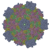
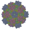
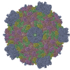
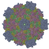
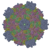
| Entry | Database: PDB / ID: 6djy | |||||||||
|---|---|---|---|---|---|---|---|---|---|---|
| Title | Fako virus | |||||||||
 Components Components |
| |||||||||
 Keywords Keywords |  VIRUS / capsid / virion / Reoviridae / T=2 VIRUS / capsid / virion / Reoviridae / T=2 | |||||||||
| Function / homology | Major capsid protein / Turret protein / Clamp protein Function and homology information Function and homology information | |||||||||
| Biological species |  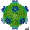 Fako virus Fako virus | |||||||||
| Method | ELECTRON MICROSCOPY / single particle reconstruction / cryo EM / Resolution: 3.9 Å | |||||||||
 Authors Authors | Kaelber, J.T. / Jiang, W. / Weaver, S.C. / Auguste, A.J. / Chiu, W. | |||||||||
| Funding support |  United States, 2items United States, 2items
| |||||||||
 Citation Citation | Journal: Structure / Year: 2020 Title: Arrangement of the Polymerase Complexes inside a Nine-Segmented dsRNA Virus. Authors: Jason T Kaelber / Wen Jiang / Scott C Weaver / Albert J Auguste / Wah Chiu / Abstract: Members of the family Reoviridae package several copies of the viral polymerase complex into their capsid to carry out replication and transcription within viral particles. Classical single-particle ...Members of the family Reoviridae package several copies of the viral polymerase complex into their capsid to carry out replication and transcription within viral particles. Classical single-particle reconstruction encounters difficulties resolving structures such as the intraparticle polymerase complex because refinement can converge to an incorrect map and because the map could depict a nonrepresentative subset of particles or an average of heterogeneous particles. Using the nine-segmented Fako virus, we tested hypotheses for the arrangement and number of polymerase complexes within the virion by measuring how well each hypothesis describes the set of cryoelectron microscopy images of individual viral particles. We find that the polymerase complex in Fako virus binds at ten possible sites despite having only nine genome segments. A single asymmetric configuration describes the arrangement of these complexes in both virions and genome-free capsids. Similarities between the arrangements of Reoviridae with 9, 10, and 11 segments indicate the generalizability of this architecture. | |||||||||
| History |
|
- Structure visualization
| Movie |
Movie viewer |
|---|---|
| Structure viewer | Molecule: MolmilJmol/JSmol |
- Downloads & links
-Download
| PDBx/mmCIF format | 6djy.cif.gz | 630.3 KB | Display | PDBx/mmCIF format |
|---|---|---|---|---|
| PDB format | pdb6djy.ent.gz | 506.5 KB | Display | PDB format |
| PDBx/mmJSON format | 6djy.json.gz | Tree view | PDBx/mmJSON format | |
| Others |  Other downloads Other downloads |
-Validation report
| Arichive directory | https://data.pdbj.org/pub/pdb/validation_reports/dj/6djyftp://data.pdbj.org/pub/pdb/validation_reports/dj/6djy | HTTPS FTP |
|---|
-Related structure data
| Related structure data | 7944MC 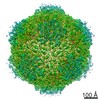 7941C 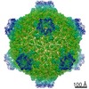 7945C 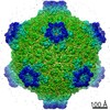 7948C 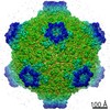 7949C 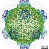 7953C 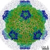 7954C C: citing same article ( M: map data used to model this data |
|---|---|
| Similar structure data |

-Links
 PDBj
PDBj- Assembly
| Deposited unit | 
|
|---|---|
| 1 | x 60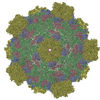
|
| 2 |
|
| 3 | x 5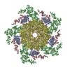
|
| 4 | x 6
|
| 5 | 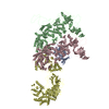
|
| Symmetry | Point symmetry: (Schoenflies symbol: I (icosahedral)) |
-Components
| #1: Protein | Mass: 39589.691 Da / Num. of mol.: 1 / Source method: isolated from a natural source / Source: (natural) Fako virus / Strain: CSW77 / References: UniProt: A0A0A0UEE5 | ||
|---|---|---|---|
| #2: Protein | Mass: 136689.766 Da / Num. of mol.: 2 / Source method: isolated from a natural source / Source: (natural) Fako virus / Strain: CSW77 / References: UniProt: A0A0A0U7Z7#3: Protein | | Mass: 121232.570 Da / Num. of mol.: 1 / Source method: isolated from a natural source / Source: (natural) Fako virus / Strain: CSW77 / References: UniProt: A0A0A0U955 |
-Experimental details
-Experiment
| Experiment | Method: ELECTRON MICROSCOPY |
|---|---|
| EM experiment | Aggregation state: PARTICLE / 3D reconstruction method: single particle reconstruction |
- Sample preparation
| Component | Name: Fako virus / Type: VIRUS / Entity ID: all / Source: NATURAL | ||||||||||||||||||||
|---|---|---|---|---|---|---|---|---|---|---|---|---|---|---|---|---|---|---|---|---|---|
| Molecular weight | Value: 43 MDa / Experimental value: NO | ||||||||||||||||||||
| Source (natural) | Organism: Fako virus / Strain: CSW77 | ||||||||||||||||||||
| Details of virus | Empty: NO / Enveloped: NO / Isolate: STRAIN / Type: VIRION | ||||||||||||||||||||
| Natural host | Organism: Culicinae | ||||||||||||||||||||
| Virus shell | Triangulation number (T number): 2 | ||||||||||||||||||||
| Buffer solution | pH: 7.8 | ||||||||||||||||||||
| Buffer component |
| ||||||||||||||||||||
| Specimen | Embedding applied: NO / Shadowing applied: NO / Staining applied: NO / Vitrification applied: YES | ||||||||||||||||||||
| Specimen support | Details: unspecified | ||||||||||||||||||||
| Vitrification | Instrument: FEI VITROBOT MARK IV / Cryogen name: ETHANE |
- Electron microscopy imaging
| Microscopy | Model: JEOL 3200FSC |
|---|---|
| Electron gun | Electron source: FIELD EMISSION GUN / Accelerating voltage: 300 kV / Illumination mode: FLOOD BEAM |
| Electron lens | Mode: BRIGHT FIELDBright-field microscopy / Cs: 4.7 mm |
| Specimen holder | Cryogen: NITROGEN / Specimen holder model: JEOL 3200FSC CRYOHOLDER |
| Image recording | Electron dose: 30 e/Å2 / Detector mode: INTEGRATING / Film or detector model: DIRECT ELECTRON DE-20 (5k x 3k) / Num. of real images: 2400 |
| EM imaging optics | Energyfilter name: In-column Omega Filter |
- Processing
| Software | Name: PHENIX / Version: dev_3366: / Classification: refinement | ||||||||||||||||||||||||
|---|---|---|---|---|---|---|---|---|---|---|---|---|---|---|---|---|---|---|---|---|---|---|---|---|---|
| EM software |
| ||||||||||||||||||||||||
| CTF correction | Type: PHASE FLIPPING AND AMPLITUDE CORRECTION | ||||||||||||||||||||||||
| Particle selection | Num. of particles selected: 10588 | ||||||||||||||||||||||||
| Symmetry | Point symmetry: I (icosahedral) | ||||||||||||||||||||||||
| 3D reconstruction | Resolution: 3.9 Å / Resolution method: FSC 0.143 CUT-OFF / Num. of particles: 5294 / Algorithm: FOURIER SPACE / Symmetry type: POINT | ||||||||||||||||||||||||
| Refine LS restraints |
|
