[English] 日本語
Yorodumi
- EMDB-5160: Atomic CryoEM Structure of a Non-Enveloped Virus Reveals How its ... -

+
Open data


ID or keywords:

Loading...

-
Basic information

Entry
Database: EMDB / ID: EMD-5160
TitleAtomic CryoEM Structure of a Non-Enveloped Virus Reveals How its Membrane Protein is Primed for Cell Entry
Map dataAquareovirus filtered to 3.2A
Sample
  • Sample: Aquareovirus
  • Virus: Aquareovirus
KeywordsAtomic CryoEM Non-enveloped Virus Membrane penetration Protein Autocleavage
Function / homology
Function and homology information


host cell surface binding / viral inner capsid / viral outer capsid / permeabilization of host organelle membrane involved in viral entry into host cell / symbiont entry into host cell via permeabilization of inner membrane / 7-methylguanosine mRNA capping / viral capsid / mRNA guanylyltransferase activity / mRNA 5'-cap (guanine-N7-)-methyltransferase activity / RNA helicase activity ...host cell surface binding / viral inner capsid / viral outer capsid / permeabilization of host organelle membrane involved in viral entry into host cell / symbiont entry into host cell via permeabilization of inner membrane / 7-methylguanosine mRNA capping / viral capsid / mRNA guanylyltransferase activity / mRNA 5'-cap (guanine-N7-)-methyltransferase activity / RNA helicase activity / RNA helicase / hydrolase activity / GTP binding / ATP binding / metal ion binding
Similarity search - Function
Mu1 membrane penetration protein, domain III / Outer capsid protein Mu1/VP4 / Mu1 membrane penetration protein, domain IV / Mu1 membrane penetration protein, domain II / Mu1/VP4 superfamily / Reovirus major virion structural protein Mu-1/Mu-1C (M2) / Sigma1/sigma2, reoviral / Reoviral Sigma1/Sigma2 family / Reovirus core-spike lambda-2 / Reovirus core-spike protein lambda-2 (L2), C-terminal ...Mu1 membrane penetration protein, domain III / Outer capsid protein Mu1/VP4 / Mu1 membrane penetration protein, domain IV / Mu1 membrane penetration protein, domain II / Mu1/VP4 superfamily / Reovirus major virion structural protein Mu-1/Mu-1C (M2) / Sigma1/sigma2, reoviral / Reoviral Sigma1/Sigma2 family / Reovirus core-spike lambda-2 / Reovirus core-spike protein lambda-2 (L2), C-terminal / Inner capsid protein lambda-1/ VP3 / C2H2-type zinc finger / zinc finger / Zinc finger C2H2 type domain profile. / Zinc finger C2H2 type domain signature. / Zinc finger C2H2-type / S-adenosyl-L-methionine-dependent methyltransferase superfamily / Immunoglobulin-like fold
Similarity search - Domain/homology
Core protein VP6 / Putative outer capsid VP4 / RNA helicase / VP1
Similarity search - Component
Biological speciesAquareovirus
Methodsingle particle reconstruction / cryo EM / Resolution: 3.3 Å
AuthorsZhang X / Jin L / Fang Q / Hui WH / Zhou ZH
Citation
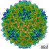
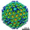
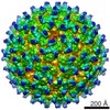
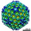
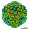
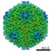
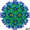
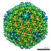
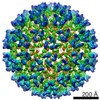
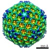
Journal: Cell / Year: 2010
Title: 3.3 A cryo-EM structure of a nonenveloped virus reveals a priming mechanism for cell entry.
Authors: Xing Zhang / Lei Jin / Qin Fang / Wong H Hui / Z Hong Zhou /
Abstract: To achieve cell entry, many nonenveloped viruses must transform from a dormant to a primed state. In contrast to the membrane fusion mechanism of enveloped viruses (e.g., influenza virus), this ...To achieve cell entry, many nonenveloped viruses must transform from a dormant to a primed state. In contrast to the membrane fusion mechanism of enveloped viruses (e.g., influenza virus), this membrane penetration mechanism is poorly understood. Here, using single-particle cryo-electron microscopy, we report a 3.3 A structure of the primed, infectious subvirion particle of aquareovirus. The density map reveals side-chain densities of all types of amino acids (except glycine), enabling construction of a full-atom model of the viral particle. Our structure and biochemical results show that priming involves autocleavage of the membrane penetration protein and suggest that Lys84 and Glu76 may facilitate this autocleavage in a nucleophilic attack. We observe a myristoyl group, covalently linked to the N terminus of the penetration protein and embedded in a hydrophobic pocket. These results suggest a well-orchestrated process of nonenveloped virus entry involving autocleavage of the penetration protein prior to exposure of its membrane-insertion finger.
History
DepositionJan 18, 2010-
Header (metadata) releaseMay 3, 2010-
Map releaseMay 3, 2010-
UpdateDec 26, 2012-
Current statusDec 26, 2012Processing site: RCSB / Status: Released

-
Structure visualization

Movie
  • Surface view with section colored by density value
  • Surface level: 0.04
  • Imaged by UCSF Chimera
  • Download
  • Surface view colored by radius
  • Surface level: 0.04
  • Imaged by UCSF Chimera
  • Download
  • Surface view with fitted model
  • Atomic models: PDB-3iyl
  • Surface level: 0.04
  • Imaged by UCSF Chimera
  • Download
  • Simplified surface model + fitted atomic model
  • Atomic modelsPDB-3iyl
  • Imaged by Jmol
  • Download
Movie viewer
Structure viewerEM map:
SurfViewMolmilJmol/JSmol
Supplemental images

Downloads & links

-
Map

FileDownload / File: emd_5160.map.gz / Format: CCP4 / Size: 1.5 GB / Type: IMAGE STORED AS FLOATING POINT NUMBER (4 BYTES)
AnnotationAquareovirus filtered to 3.2A
Voxel sizeX=Y=Z: 1.1 Å
Density
Contour LevelBy EMDB: 0.05 / Movie #1: 0.04
Minimum - Maximum-0.19343019 - 0.30496365
Average (Standard dev.)-0.00054387 (±0.02425474)
SymmetrySpace group: 1
Details

EMDB XML:

Map geometry
Axis orderXYZ
Origin-370-370-370
Dimensions740740740
Spacing740740740
CellA=B=C: 814.0 Å
α=β=γ: 90.0 °

CCP4 map header:

modeImage stored as Reals
Å/pix. X/Y/Z1.11.11.1
M x/y/z740740740
origin x/y/z0.0000.0000.000
length x/y/z814.000814.000814.000
α/β/γ90.00090.00090.000
start NX/NY/NZ-34-26-72
NX/NY/NZ6953145
MAP C/R/S123
start NC/NR/NS-370-370-370
NC/NR/NS740740740
D min/max/mean-0.1930.305-0.001

-
Supplemental data

-
Segmentation: averaged VP5 protein at 3.2a.

Annotationaveraged VP5 protein at 3.2a.
Fileemd_5160_msk_1.map
Projections & Slices
AxesZYX

Projections

Slices (1/2)
Density Histograms

-
Sample components

-
Entire : Aquareovirus

EntireName: Aquareovirus
Components
  • Sample: Aquareovirus
  • Virus: Aquareovirus

-
Supramolecule #1000: Aquareovirus

SupramoleculeName: Aquareovirus / type: sample / ID: 1000 / Details: The sample was monodisperse / Number unique components: 4
Molecular weightExperimental: 72 MDa / Theoretical: 72 MDa

-
Supramolecule #1: Aquareovirus

SupramoleculeName: Aquareovirus / type: virus / ID: 1 / Name.synonym: GCRV / NCBI-ID: 10979 / Sci species name: Aquareovirus / Database: NCBI / Virus type: OTHER / Virus isolate: STRAIN / Virus enveloped: No / Virus empty: No / Syn species name: GCRV
Host (natural)synonym: VERTEBRATES
Molecular weightExperimental: 72 MDa / Theoretical: 72 MDa
Virus shellShell ID: 1 / Name: ISVP / Diameter: 800 Å / T number (triangulation number): 13

-
Experimental details

-
Structure determination

Methodcryo EM
Processingsingle particle reconstruction
Aggregation stateparticle

-
Sample preparation

BufferpH: 7.5 / Details: 10mM PBS Buffer
GridDetails: 400 mesh quantifoil 1.2/1.3
VitrificationCryogen name: METHANE / Chamber humidity: 100 % / Chamber temperature: 90 K / Instrument: OTHER / Details: Vitrification instrument: FEI Vitrobot / Method: Blot for 7-9 seconds before plunging

-
Electron microscopy

MicroscopeFEI TITAN KRIOS
Electron beamAcceleration voltage: 300 kV / Electron source: FIELD EMISSION GUN
Electron opticsCalibrated magnification: 57700 / Illumination mode: FLOOD BEAM / Imaging mode: BRIGHT FIELDBright-field microscopy / Cs: 2.7 mm / Nominal defocus max: 2.7 µm / Nominal defocus min: 0.4 µm / Nominal magnification: 59000
Sample stageSpecimen holder: Eucentric / Specimen holder model: OTHER
TemperatureMin: 90 K / Average: 90 K
Alignment procedureLegacy - Astigmatism: objective lens astigmatism was corrected at 250,000 times magnification
DateMar 1, 2009
Image recordingCategory: CCD / Film or detector model: KODAK SO-163 FILM / Digitization - Sampling interval: 6.35 µm / Number real images: 700 / Average electron dose: 25 e/Å2 / Bits/pixel: 16
Experimental equipment
Model: Titan Krios / Image courtesy: FEI Company

-
Image processing

CTF correctionDetails: Each particle
Final angle assignmentDetails: Frealign IMIRS
Final reconstructionAlgorithm: OTHER / Resolution.type: BY AUTHOR / Resolution: 3.3 Å / Resolution method: FSC 0.143 CUT-OFF / Software - Name: Frealign IMIRS / Number images used: 18464

+
About Yorodumi

-
News

-
Feb 9, 2022. New format data for meta-information of EMDB entries

New format data for meta-information of EMDB entries

  • Version 3 of the EMDB header file is now the official format.
  • The previous official version 1.9 will be removed from the archive.

Related info.:EMDB header

External links:wwPDB to switch to version 3 of the EMDB data model

-
Aug 12, 2020. Covid-19 info

Covid-19 info

URL: https://pdbj.org/emnavi/covid19.php

New page: Covid-19 featured information page in EM Navigator.

Related info.:Covid-19 info / Mar 5, 2020. Novel coronavirus structure data

+
Mar 5, 2020. Novel coronavirus structure data

Novel coronavirus structure data

Related info.:Yorodumi Speices / Aug 12, 2020. Covid-19 info

External links:COVID-19 featured content - PDBj / Molecule of the Month (242):Coronavirus Proteases

+
Jan 31, 2019. EMDB accession codes are about to change! (news from PDBe EMDB page)

EMDB accession codes are about to change! (news from PDBe EMDB page)

  • The allocation of 4 digits for EMDB accession codes will soon come to an end. Whilst these codes will remain in use, new EMDB accession codes will include an additional digit and will expand incrementally as the available range of codes is exhausted. The current 4-digit format prefixed with “EMD-” (i.e. EMD-XXXX) will advance to a 5-digit format (i.e. EMD-XXXXX), and so on. It is currently estimated that the 4-digit codes will be depleted around Spring 2019, at which point the 5-digit format will come into force.
  • The EM Navigator/Yorodumi systems omit the EMD- prefix.

Related info.:Q: What is EMD? / ID/Accession-code notation in Yorodumi/EM Navigator

External links:EMDB Accession Codes are Changing Soon! / Contact to PDBj

+
Jul 12, 2017. Major update of PDB

Major update of PDB

  • wwPDB released updated PDB data conforming to the new PDBx/mmCIF dictionary.
  • This is a major update changing the version number from 4 to 5, and with Remediation, in which all the entries are updated.
  • In this update, many items about electron microscopy experimental information are reorganized (e.g. em_software).
  • Now, EM Navigator and Yorodumi are based on the updated data.

External links:wwPDB Remediation / Enriched Model Files Conforming to OneDep Data Standards Now Available in the PDB FTP Archive

-
Yorodumi

Thousand views of thousand structures

  • Yorodumi is a browser for structure data from EMDB, PDB, SASBDB, etc.
  • This page is also the successor to EM Navigator detail page, and also detail information page/front-end page for Omokage search.
  • The word "yorodu" (or yorozu) is an old Japanese word meaning "ten thousand". "mi" (miru) is to see.

Related info.:EMDB / PDB / SASBDB / Comparison of 3 databanks / Yorodumi Search / Aug 31, 2016. New EM Navigator & Yorodumi / Yorodumi Papers / Jmol/JSmol / Function and homology information / Changes in new EM Navigator and Yorodumi

Read more