 Movie
Movie Controller

[English] 日本語
 Yorodumi
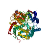
- PDB-3hxj: Crystal Structure of Pyrrolo-quinoline quinone (PQQ_DH) from Meth... -
+ Open data

- Basic information
| Entry | Database: PDB / ID: 3hxj | ||||||
|---|---|---|---|---|---|---|---|
| Title | Crystal Structure of Pyrrolo-quinoline quinone (PQQ_DH) from Methanococcus maripaludis, Northeast Structural Genomics Consortium Target MrR86 | ||||||
 Components Components | Pyrrolo-quinoline quinone | ||||||
 Keywords Keywords |  OXIDOREDUCTASE / All beta protein. Incomplete 8-blade beta-propeller. / Structural Genomics / PSI-2 / Protein Structure Initiative / Northeast Structural Genomics Consortium / NESG OXIDOREDUCTASE / All beta protein. Incomplete 8-blade beta-propeller. / Structural Genomics / PSI-2 / Protein Structure Initiative / Northeast Structural Genomics Consortium / NESG | ||||||
| Function / homology | : / PQQ-like domain / Pyrrolo-quinoline quinone repeat / PQQ-like domain / Pyrrolo-quinoline quinone beta-propeller repeat / beta-propeller repeat / Quinoprotein alcohol dehydrogenase-like superfamily / WD40/YVTN repeat-like-containing domain superfamily / Pyrrolo-quinoline quinone Function and homology information Function and homology information | ||||||
| Biological species |  Methanococcus maripaludis (archaea) Methanococcus maripaludis (archaea) | ||||||
| Method | X-RAY DIFFRACTION / SYNCHROTRON / SAD / Resolution: 2 Å | ||||||
 Authors Authors | Forouhar, F. / Chen, Y. / Seetharaman, J. / Sahdev, S. / Xiao, R. / Ciccosanti, C. / Foote, E.L. / Zhao, L. / Everett, J.K. / Nair, R. ...Forouhar, F. / Chen, Y. / Seetharaman, J. / Sahdev, S. / Xiao, R. / Ciccosanti, C. / Foote, E.L. / Zhao, L. / Everett, J.K. / Nair, R. / Acton, T.B. / Rost, B. / Montelione, G.T. / Hunt, J.F. / Tong, L. / Northeast Structural Genomics Consortium (NESG) | ||||||
 Citation Citation | Journal: To be Published Title: Northeast Structural Genomics Consortium Target MrR86 Authors: Forouhar, F. / Chen, Y. / Seetharaman, J. / Sahdev, S. / Xiao, R. / Ciccosanti, C. / Foote, E.L. / Zhao, L. / Everett, J.K. / Nair, R. / Acton, T.B. / Rost, B. / Montelione, G.T. / Hunt, J.F. / Tong, L. | ||||||
| History |
|
- Structure visualization
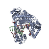
| Structure viewer | Molecule: MolmilJmol/JSmol |
|---|
- Downloads & links
-Download
| PDBx/mmCIF format | 3hxj.cif.gz | 255.5 KB | Display | PDBx/mmCIF format |
|---|---|---|---|---|
| PDB format | pdb3hxj.ent.gz | 213.6 KB | Display | PDB format |
| PDBx/mmJSON format | 3hxj.json.gz | Tree view | PDBx/mmJSON format | |
| Others |  Other downloads Other downloads |
-Validation report
| Arichive directory | https://data.pdbj.org/pub/pdb/validation_reports/hx/3hxjftp://data.pdbj.org/pub/pdb/validation_reports/hx/3hxj | HTTPS FTP |
|---|
-Related structure data
| Similar structure data | |
|---|---|
| Other databases |

-Links
 PDBj
PDBj- Assembly
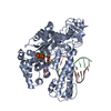
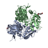
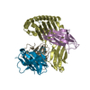
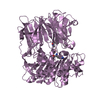
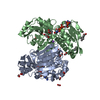
| Deposited unit | 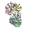
| ||||||||
|---|---|---|---|---|---|---|---|---|---|
| 1 | 
| ||||||||
| 2 | 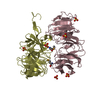
| ||||||||
| Unit cell |
|
-Components
| #1: Protein | Mass: 37285.074 Da / Num. of mol.: 4 / Mutation: L115M,P124T Source method: isolated from a genetically manipulated source Source: (gene. exp.) Methanococcus maripaludis (archaea) / Strain: C7 / Gene: MmarC7 1297, MmarC7_1297 / Plasmid: BL21 / Production host:  Escherichia coli (E. coli) / Strain (production host): BL21(DE3)+ Magic / References: UniProt: A6VIT4 Escherichia coli (E. coli) / Strain (production host): BL21(DE3)+ Magic / References: UniProt: A6VIT4#2: Chemical | ChemComp-SO4 / Sulfate  Mass: 96.063 Da / Num. of mol.: 7 / Source method: obtained synthetically / Formula: SO4 Mass: 96.063 Da / Num. of mol.: 7 / Source method: obtained synthetically / Formula: SO4#3: Water | ChemComp-HOH / | Water Mass: 18.015 Da / Num. of mol.: 669 / Source method: isolated from a natural source / Formula: H2O Mass: 18.015 Da / Num. of mol.: 669 / Source method: isolated from a natural source / Formula: H2OSequence details | IN ORDER TO DETERMINE THE CRYSTAL STRUCTURE OF THIS PROTEIN BY SAD PHASING USING SE-MET SIGNAL, LEU ...IN ORDER TO DETERMINE THE CRYSTAL STRUCTURE OF THIS PROTEIN BY SAD PHASING USING SE-MET SIGNAL, LEU 115 WAS MUTATED TO MET. RESIDUE 124 IS THR, NOT PRO BASED ON THE CURRENT CRYSTAL STRUCTURE, FOR THE ELECTRON DENSITY FOR THR IS UNAMBIGUOU | |
|---|
-Experimental details
-Experiment
| Experiment | Method: X-RAY DIFFRACTION / Number of used crystals: 1 |
|---|
- Sample preparation
| Crystal | Density Matthews: 2.26 Å3/Da / Density % sol: 45.51 % |
|---|---|
| Crystal grow | Temperature: 291 K / Method: microbatch, under oil / pH: 5.5 Details: Protein solution: 100mM NaCl, 5mM DTT, 0.02% NaN3, 10mM Tris-HCl (pH 7.5), Reservoir solution: 0.1 M Bis-Tris (pH 5.5), 25% PEG3350, and 0.2 M Lithium sulfate, microbatch, under oil, temperature 291K |
-Data collection
| Diffraction | Mean temperature: 100 K |
|---|---|
| Diffraction source | Source: SYNCHROTRON / Site: SSRL  / Beamline: BL9-2 / Wavelength: 0.97905 Å / Beamline: BL9-2 / Wavelength: 0.97905 Å |
| Detector | Type: MARMOSAIC 325 mm CCD / Detector: CCD / Date: May 31, 2009 / Details: mirrors |
| Radiation | Monochromator: Si 111 CHANNEL / Protocol: SINGLE WAVELENGTH / Monochromatic (M) / Laue (L): M / Scattering type: x-ray |
| Radiation wavelength | Wavelength: 0.97905 Å / Relative weight: 1 |
| Reflection | Resolution: 2→30 Å / Num. all: 176293 / Num. obs: 173972 / % possible obs: 98.7 % / Observed criterion σ(F): 0 / Observed criterion σ(I): 0 / Redundancy: 3.6 % / Biso Wilson estimate: 7.9 Å2 / Rmerge(I) obs: 0.067 / Rsym value: 0.058 / Net I/σ(I): 17.5 |
| Reflection shell | Resolution: 2→2.07 Å / Redundancy: 2.4 % / Rmerge(I) obs: 0.281 / Mean I/σ(I) obs: 3 / Num. unique all: 17652 / Rsym value: 0.241 / % possible all: 89.2 |
- Processing
| Software |
| ||||||||||||||||||||||||||||||||
|---|---|---|---|---|---|---|---|---|---|---|---|---|---|---|---|---|---|---|---|---|---|---|---|---|---|---|---|---|---|---|---|---|---|
| Refinement | Method to determine structure: SAD / Resolution: 2→19.98 Å / Rfactor Rfree error: 0.003 / Data cutoff high absF: 219394.594 / Data cutoff low absF: 0 / Isotropic thermal model: RESTRAINED / Cross valid method: THROUGHOUT / σ(F): 2 / σ(I): 2 / Stereochemistry target values: Engh & Huber
| ||||||||||||||||||||||||||||||||
| Solvent computation | Solvent model: FLAT MODEL / Bsol: 54.696 Å2 / ksol: 0.4 e/Å3 | ||||||||||||||||||||||||||||||||
| Displacement parameters | Biso mean: 26.5 Å2
| ||||||||||||||||||||||||||||||||
| Refine analyze |
| ||||||||||||||||||||||||||||||||
| Refinement step | Cycle: LAST / Resolution: 2→19.98 Å
| ||||||||||||||||||||||||||||||||
| Refine LS restraints |
| ||||||||||||||||||||||||||||||||
| LS refinement shell | Resolution: 2→2.07 Å / Rfactor Rfree error: 0.012 / Total num. of bins used: 10
|
