[English] 日本語
Yorodumi
- PDB-3qx3: Human topoisomerase IIbeta in complex with DNA and etoposide -

+
Open data


ID or keywords:

Loading...

-
Basic information

Entry
Database: PDB / ID: 3qx3
TitleHuman topoisomerase IIbeta in complex with DNA and etoposide
Components
  • DNA (5'-D(P*AP*GP*CP*CP*GP*AP*GP*C)-3')
  • DNA (5'-D(P*TP*GP*CP*AP*GP*CP*TP*CP*GP*GP*CP*T)-3')
  • DNA topoisomerase 2-beta
KeywordsISOMERASE/DNA/ISOMERASE INHIBITOR / TOPRIM domain / winged-helix domain / coiled-coil domain / DNA binding and cleavage / nucleus / ISOMERASE-DNA-ISOMERASE INHIBITOR complex
Function / homology
Function and homology information


positive regulation of single stranded viral RNA replication via double stranded DNA intermediate / sister chromatid segregation / resolution of meiotic recombination intermediates / DNA topoisomerase type II (double strand cut, ATP-hydrolyzing) activity / DNA topoisomerase (ATP-hydrolysing) / cellular response to ATP / positive regulation of double-strand break repair via nonhomologous end joining / DNA topological change / SUMOylation of DNA replication proteins / ribonucleoprotein complex binding ...positive regulation of single stranded viral RNA replication via double stranded DNA intermediate / sister chromatid segregation / resolution of meiotic recombination intermediates / DNA topoisomerase type II (double strand cut, ATP-hydrolyzing) activity / DNA topoisomerase (ATP-hydrolysing) / cellular response to ATP / positive regulation of double-strand break repair via nonhomologous end joining / DNA topological change / SUMOylation of DNA replication proteins / ribonucleoprotein complex binding / forebrain development / axonogenesis / B cell differentiation / neuron migration / cellular response to hydrogen peroxide / cellular senescence / ribonucleoprotein complex / chromatin binding / nucleolus / DNA binding / nucleoplasm / ATP binding / metal ion binding / nucleus / cytosol
Similarity search - Function
Topoisomerase II; domain 5 / Topoisomerase II, domain 5 / DTHCT / DTHCT (NUC029) region / Topoisomerase, domain 3 / Topoisomerase; domain 3 / DNA topoisomerase 2, TOPRIM domain / Rossmann fold - #670 / Gyrase A; domain 2 - #40 / DNA topoisomerase II, eukaryotic-type ...Topoisomerase II; domain 5 / Topoisomerase II, domain 5 / DTHCT / DTHCT (NUC029) region / Topoisomerase, domain 3 / Topoisomerase; domain 3 / DNA topoisomerase 2, TOPRIM domain / Rossmann fold - #670 / Gyrase A; domain 2 - #40 / DNA topoisomerase II, eukaryotic-type / C-terminal associated domain of TOPRIM / C-terminal associated domain of TOPRIM / Topoisomerase (Topo) IIA-type catalytic domain profile. / DNA topoisomerase, type IIA, alpha-helical domain superfamily / DNA topoisomerase, type IIA, domain A / DNA topoisomerase, type IIA, domain A, alpha-beta / DNA gyrase/topoisomerase IV, subunit A / DNA Topoisomerase IV / DNA topoisomerase, type IIA, subunit B, domain 2 / DNA gyrase B / DNA topoisomerase, type IIA / DNA topoisomerase, type IIA, conserved site / DNA topoisomerase II signature. / TopoisomeraseII / DNA topoisomerase, type IIA, subunit B, C-terminal / Toprim domain / DNA topoisomerase, type IIA-like domain superfamily / Toprim domain profile. / TOPRIM domain / Gyrase A; domain 2 / Histidine kinase-, DNA gyrase B-, and HSP90-like ATPase / Histidine kinase/HSP90-like ATPase / Histidine kinase/HSP90-like ATPase superfamily / Ribosomal protein S5 domain 2-type fold, subgroup / Ribosomal protein S5 domain 2-type fold / Alpha-Beta Complex / Rossmann fold / 2-Layer Sandwich / Orthogonal Bundle / 3-Layer(aba) Sandwich / Mainly Alpha / Alpha Beta
Similarity search - Domain/homology
Chem-EVP / DNA / DNA (> 10) / DNA topoisomerase 2-beta
Similarity search - Component
Biological speciesHomo sapiens (human)
MethodX-RAY DIFFRACTION / SYNCHROTRON / MOLECULAR REPLACEMENT / Resolution: 2.162 Å
AuthorsWu, C.C. / Li, T.K. / Farh, L. / Lin, L.Y. / Lin, T.S. / Yu, Y.J. / Yen, T.J. / Chiang, C.W. / Chan, N.L.
CitationJournal: Science / Year: 2011
Title: Structural basis of type II topoisomerase inhibition by the anticancer drug etoposide
Authors: Wu, C.C. / Li, T.K. / Farh, L. / Lin, L.Y. / Lin, T.S. / Yu, Y.J. / Yen, T.J. / Chiang, C.W. / Chan, N.L.
History
DepositionMar 1, 2011Deposition site: RCSB / Processing site: PDBJ
Revision 1.0Jul 6, 2011Provider: repository / Type: Initial release
Revision 1.1Jul 13, 2011Group: Version format compliance
Revision 1.2Jan 25, 2012Group: Database references
Revision 1.3Nov 1, 2023Group: Data collection / Database references ...Data collection / Database references / Derived calculations / Refinement description / Structure summary
Category: chem_comp / chem_comp_atom ...chem_comp / chem_comp_atom / chem_comp_bond / database_2 / entity / pdbx_entity_nonpoly / pdbx_initial_refinement_model / pdbx_struct_conn_angle / struct_conn / struct_conn_type / struct_ref_seq_dif / struct_site
Item: _chem_comp.name / _database_2.pdbx_DOI ..._chem_comp.name / _database_2.pdbx_DOI / _database_2.pdbx_database_accession / _entity.pdbx_description / _pdbx_entity_nonpoly.name / _pdbx_struct_conn_angle.ptnr1_auth_asym_id / _pdbx_struct_conn_angle.ptnr1_auth_comp_id / _pdbx_struct_conn_angle.ptnr1_auth_seq_id / _pdbx_struct_conn_angle.ptnr1_label_asym_id / _pdbx_struct_conn_angle.ptnr1_label_atom_id / _pdbx_struct_conn_angle.ptnr1_label_comp_id / _pdbx_struct_conn_angle.ptnr1_label_seq_id / _pdbx_struct_conn_angle.ptnr2_auth_asym_id / _pdbx_struct_conn_angle.ptnr2_auth_seq_id / _pdbx_struct_conn_angle.ptnr2_label_asym_id / _pdbx_struct_conn_angle.ptnr3_auth_asym_id / _pdbx_struct_conn_angle.ptnr3_auth_comp_id / _pdbx_struct_conn_angle.ptnr3_auth_seq_id / _pdbx_struct_conn_angle.ptnr3_label_asym_id / _pdbx_struct_conn_angle.ptnr3_label_atom_id / _pdbx_struct_conn_angle.ptnr3_label_comp_id / _pdbx_struct_conn_angle.ptnr3_label_seq_id / _pdbx_struct_conn_angle.value / _struct_conn.conn_type_id / _struct_conn.id / _struct_conn.pdbx_dist_value / _struct_conn.pdbx_leaving_atom_flag / _struct_conn.ptnr1_auth_asym_id / _struct_conn.ptnr1_auth_comp_id / _struct_conn.ptnr1_auth_seq_id / _struct_conn.ptnr1_label_asym_id / _struct_conn.ptnr1_label_atom_id / _struct_conn.ptnr1_label_comp_id / _struct_conn.ptnr1_label_seq_id / _struct_conn.ptnr2_auth_asym_id / _struct_conn.ptnr2_auth_comp_id / _struct_conn.ptnr2_auth_seq_id / _struct_conn.ptnr2_label_asym_id / _struct_conn.ptnr2_label_atom_id / _struct_conn.ptnr2_label_comp_id / _struct_conn.ptnr2_label_seq_id / _struct_conn_type.id / _struct_ref_seq_dif.details / _struct_site.pdbx_auth_asym_id / _struct_site.pdbx_auth_comp_id / _struct_site.pdbx_auth_seq_id

-
Structure visualization

Structure viewerMolecule:
MolmilJmol/JSmol

Downloads & links

-
Assembly

Deposited unit
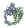
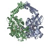
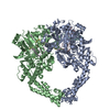
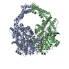
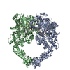
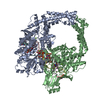
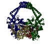
A: DNA topoisomerase 2-beta
B: DNA topoisomerase 2-beta
C: DNA (5'-D(P*AP*GP*CP*CP*GP*AP*GP*C)-3')
D: DNA (5'-D(P*TP*GP*CP*AP*GP*CP*TP*CP*GP*GP*CP*T)-3')
E: DNA (5'-D(P*AP*GP*CP*CP*GP*AP*GP*C)-3')
F: DNA (5'-D(P*TP*GP*CP*AP*GP*CP*TP*CP*GP*GP*CP*T)-3')
hetero molecules


Theoretical massNumber of molelcules
Total (without water)197,36214
Polymers196,0396
Non-polymers1,3238
Water18,8621047
1


  • Idetical with deposited unit
  • defined by author&software
TypeNameSymmetry operationNumber
identity operation1_555x,y,z1
Buried area12490 Å2
ΔGint-70 kcal/mol
Surface area64430 Å2
MethodPISA
Unit cell
Length a, b, c (Å)80.149, 176.801, 94.067
Angle α, β, γ (deg.)90.00, 111.58, 90.00
Int Tables number4
Space group name H-MP1211

-
Components

-
Protein , 1 types, 2 molecules AB

#1: Protein DNA topoisomerase 2-beta / DNA topoisomerase II / beta isozyme


Mass: 91928.367 Da / Num. of mol.: 2 / Fragment: hTOP2beta cleavage core, UNP residues 450-1206
Source method: isolated from a genetically manipulated source
Source: (gene. exp.) Homo sapiens (human) / Gene: TOP2B / Plasmid: pET51b / Production host: Escherichia coli (E. coli) / Strain (production host): BL21star(DE3) / References: UniProt: Q02880, EC: 5.99.1.3

-
DNA chain , 2 types, 4 molecules CEDF

#2: DNA chain DNA (5'-D(P*AP*GP*CP*CP*GP*AP*GP*C)-3')


Mass: 2436.619 Da / Num. of mol.: 2 / Source method: obtained synthetically
Details: The oligonucleotide sequence 5'-AGCCGAGCTGCAGCTCGGCT-3' was purchased from VIOGENE.
#3: DNA chain DNA (5'-D(P*TP*GP*CP*AP*GP*CP*TP*CP*GP*GP*CP*T)-3')


Mass: 3654.378 Da / Num. of mol.: 2 / Source method: obtained synthetically
Details: The oligonucleotide sequence 5'-AGCCGAGCTGCAGCTCGGCT-3' was purchased from VIOGENE.

-
Non-polymers , 3 types, 1055 molecules

#4: Chemical ChemComp-EVP / (5S,5aR,8aR,9R)-9-(4-hydroxy-3,5-dimethoxyphenyl)-8-oxo-5,5a,6,8,8a,9-hexahydrofuro[3',4':6,7]naphtho[2,3-d][1,3]dioxol -5-yl 4,6-O-[(1R)-ethylidene]-beta-D-glucopyranoside / Etoposide / VP-16 / Etoposide


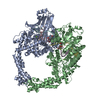
Mass: 588.557 Da / Num. of mol.: 2 / Source method: obtained synthetically / Formula: C29H32O13 / Comment: medication, chemotherapy*YM
#5: Chemical
ChemComp-MG / MAGNESIUM ION


Mass: 24.305 Da / Num. of mol.: 6 / Source method: obtained synthetically / Formula: Mg
#6: Water ChemComp-HOH / water / Water


Mass: 18.015 Da / Num. of mol.: 1047 / Source method: isolated from a natural source / Formula: H2O

-
Details

Sequence detailsTHE SEQUENCE OF DNA USED IN THE EXPERIMENT WAS 5'-AGCCGAGCTGCAGCTCGGCT-3'. AND THE DNAS WERE ...THE SEQUENCE OF DNA USED IN THE EXPERIMENT WAS 5'-AGCCGAGCTGCAGCTCGGCT-3'. AND THE DNAS WERE CLEAVED INTO TWO PIECES (TOTALLY 4 STRANDS, DESIGNATED AS CHAIN C, D, E AND F) DURING EXPERIMENT.

-
Experimental details

-
Experiment

ExperimentMethod: X-RAY DIFFRACTION / Number of used crystals: 1

-
Sample preparation

CrystalDensity Matthews: 3.16 Å3/Da / Density % sol: 61.09 %
Crystal growTemperature: 277 K / Method: vapor diffusion, hanging drop / pH: 5.8
Details: 0.1M magnesium acetate, 50mM 2-(N-morpholino)ethanesulfonic acid pH 5.8, 22% 2-methyl-2, 4-pentanediol (MPT), VAPOR DIFFUSION, HANGING DROP, temperature 277K

-
Data collection

DiffractionMean temperature: 100 K
Diffraction sourceSource: SYNCHROTRON / Site: SPring-8 / Beamline: BL12B2 / Wavelength: 0.9 Å
DetectorType: ADSC QUANTUM 210 / Detector: CCD / Date: Dec 2, 2010
RadiationMonochromator: Fixed-Exit Double Crystal Monochromator (Crystal type Si(1 1 1))
Protocol: SINGLE WAVELENGTH / Monochromatic (M) / Laue (L): M / Scattering type: x-ray
Radiation wavelengthWavelength: 0.9 Å / Relative weight: 1
ReflectionResolution: 2.16→30 Å / Num. all: 123847 / Num. obs: 129411 / % possible obs: 95.7 % / Observed criterion σ(F): 1 / Observed criterion σ(I): 1 / Redundancy: 2.6 % / Biso Wilson estimate: 32.09 Å2 / Rsym value: 0.06 / Net I/σ(I): 16.9
Reflection shellResolution: 2.16→2.2 Å / Redundancy: 2.3 % / Rmerge(I) obs: 0.472 / Mean I/σ(I) obs: 2.1 / Rsym value: 0.472 / % possible all: 98.2

-
Processing

Software
NameVersionClassification
Blu-Icedata collection
PHENIX(phenix.automr: 1.6.4_486)model building
PHENIX(phenix.refine: 1.6.4_486)refinement
HKL-2000data reduction
HKL-2000data scaling
PHENIX1.6.4_486phasing
RefinementMethod to determine structure: MOLECULAR REPLACEMENT
Starting model: PDB ENTRY 3L4K
Resolution: 2.162→29.543 Å / FOM work R set: 0.8652 / SU ML: 0.25 / σ(F): 0 / Phase error: 21.2 / Stereochemistry target values: ML
RfactorNum. reflection% reflection
Rfree0.2072 5917 5.03 %
Rwork0.1682 --
obs0.1701 117720 90.82 %
all-129619 -
Solvent computationShrinkage radii: 0.95 Å / VDW probe radii: 1.2 Å / Solvent model: FLAT BULK SOLVENT MODEL / Bsol: 40 Å2 / ksol: 0.317 e/Å3
Displacement parametersBiso mean: 37.0272 Å2
Baniso -1Baniso -2Baniso -3
1-6.3155 Å2-0 Å25.9953 Å2
2--1.0717 Å2-0 Å2
3---3.9119 Å2
Refinement stepCycle: LAST / Resolution: 2.162→29.543 Å
ProteinNucleic acidLigandSolventTotal
Num. atoms10826 820 90 1047 12783
Refine LS restraints
Refine-IDTypeDev idealNumber
X-RAY DIFFRACTIONf_bond_d0.00712326
X-RAY DIFFRACTIONf_angle_d1.05816825
X-RAY DIFFRACTIONf_dihedral_angle_d15.4424791
X-RAY DIFFRACTIONf_chiral_restr0.0691857
X-RAY DIFFRACTIONf_plane_restr0.0042017
LS refinement shell
Resolution (Å)Rfactor RfreeNum. reflection RfreeRfactor RworkNum. reflection RworkRefine-ID% reflection obs (%)
2.1619-2.23910.28825720.232510113X-RAY DIFFRACTION82
2.2391-2.32880.26895370.209710652X-RAY DIFFRACTION87
2.3288-2.43470.24825760.194511012X-RAY DIFFRACTION90
2.4347-2.5630.23165760.190211223X-RAY DIFFRACTION91
2.563-2.72350.23575740.178111420X-RAY DIFFRACTION93
2.7235-2.93360.21316300.171211577X-RAY DIFFRACTION94
2.9336-3.22850.21816520.165611605X-RAY DIFFRACTION95
3.2285-3.69490.18955940.151311700X-RAY DIFFRACTION95
3.6949-4.65220.16866250.139711317X-RAY DIFFRACTION92
4.6522-29.54620.19825810.172611184X-RAY DIFFRACTION90

+
About Yorodumi

-
News

-
Feb 9, 2022. New format data for meta-information of EMDB entries

New format data for meta-information of EMDB entries

  • Version 3 of the EMDB header file is now the official format.
  • The previous official version 1.9 will be removed from the archive.

Related info.:EMDB header

External links:wwPDB to switch to version 3 of the EMDB data model

-
Aug 12, 2020. Covid-19 info

Covid-19 info

URL: https://pdbj.org/emnavi/covid19.php

New page: Covid-19 featured information page in EM Navigator.

Related info.:Covid-19 info / Mar 5, 2020. Novel coronavirus structure data

+
Mar 5, 2020. Novel coronavirus structure data

Novel coronavirus structure data

Related info.:Yorodumi Speices / Aug 12, 2020. Covid-19 info

External links:COVID-19 featured content - PDBj / Molecule of the Month (242):Coronavirus Proteases

+
Jan 31, 2019. EMDB accession codes are about to change! (news from PDBe EMDB page)

EMDB accession codes are about to change! (news from PDBe EMDB page)

  • The allocation of 4 digits for EMDB accession codes will soon come to an end. Whilst these codes will remain in use, new EMDB accession codes will include an additional digit and will expand incrementally as the available range of codes is exhausted. The current 4-digit format prefixed with “EMD-” (i.e. EMD-XXXX) will advance to a 5-digit format (i.e. EMD-XXXXX), and so on. It is currently estimated that the 4-digit codes will be depleted around Spring 2019, at which point the 5-digit format will come into force.
  • The EM Navigator/Yorodumi systems omit the EMD- prefix.

Related info.:Q: What is EMD? / ID/Accession-code notation in Yorodumi/EM Navigator

External links:EMDB Accession Codes are Changing Soon! / Contact to PDBj

+
Jul 12, 2017. Major update of PDB

Major update of PDB

  • wwPDB released updated PDB data conforming to the new PDBx/mmCIF dictionary.
  • This is a major update changing the version number from 4 to 5, and with Remediation, in which all the entries are updated.
  • In this update, many items about electron microscopy experimental information are reorganized (e.g. em_software).
  • Now, EM Navigator and Yorodumi are based on the updated data.

External links:wwPDB Remediation / Enriched Model Files Conforming to OneDep Data Standards Now Available in the PDB FTP Archive

-
Yorodumi

Thousand views of thousand structures

  • Yorodumi is a browser for structure data from EMDB, PDB, SASBDB, etc.
  • This page is also the successor to EM Navigator detail page, and also detail information page/front-end page for Omokage search.
  • The word "yorodu" (or yorozu) is an old Japanese word meaning "ten thousand". "mi" (miru) is to see.

Related info.:EMDB / PDB / SASBDB / Comparison of 3 databanks / Yorodumi Search / Aug 31, 2016. New EM Navigator & Yorodumi / Yorodumi Papers / Jmol/JSmol / Function and homology information / Changes in new EM Navigator and Yorodumi

Read more