 ムービー
ムービー コントローラー


+ データを開く

- 基本情報
基本情報
| 登録情報 | 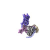 | ||||||||||||||||||
|---|---|---|---|---|---|---|---|---|---|---|---|---|---|---|---|---|---|---|---|
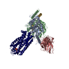
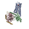
| タイトル | Cryo-EM structure of the purinergic receptor P2Y1R in complex with 2MeSADP and G11 | ||||||||||||||||||
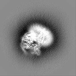
 マップデータ マップデータ | |||||||||||||||||||
 試料 試料 |
| ||||||||||||||||||
 キーワード キーワード |  G protein-coupled receptor (Gタンパク質共役受容体) / purinergic receptor / P2Y1R / ligand binding (リガンド) / signal transduction (シグナル伝達) / MEMBRANE PROTEIN (膜タンパク質) G protein-coupled receptor (Gタンパク質共役受容体) / purinergic receptor / P2Y1R / ligand binding (リガンド) / signal transduction (シグナル伝達) / MEMBRANE PROTEIN (膜タンパク質) | ||||||||||||||||||
| 機能・相同性 |  機能・相同性情報 機能・相同性情報G protein-coupled ATP receptor activity / relaxation of muscle / A1 adenosine receptor binding / G protein-coupled ADP receptor activity / P2Y受容体 / cellular response to purine-containing compound / G protein-coupled purinergic nucleotide receptor activity / positive regulation of inositol trisphosphate biosynthetic process / positive regulation of monoatomic ion transport / positive regulation of penile erection ...G protein-coupled ATP receptor activity / relaxation of muscle / A1 adenosine receptor binding / G protein-coupled ADP receptor activity / P2Y受容体 / cellular response to purine-containing compound / G protein-coupled purinergic nucleotide receptor activity / positive regulation of inositol trisphosphate biosynthetic process / positive regulation of monoatomic ion transport / positive regulation of penile erection / negative regulation of norepinephrine secretion / glial cell migration / regulation of presynaptic cytosolic calcium ion concentration / G protein-coupled adenosine receptor signaling pathway / positive regulation of hormone secretion / signal transduction involved in regulation of gene expression / response to growth factor / cellular response to ATP / regulation of synaptic vesicle exocytosis / eating behavior / monoatomic ion transport / presynaptic active zone membrane / response to mechanical stimulus / adenylate cyclase-inhibiting G protein-coupled receptor signaling pathway / blood vessel diameter maintenance / protein localization to plasma membrane / establishment of localization in cell / Olfactory Signaling Pathway / Activation of the phototransduction cascade / G beta:gamma signalling through PLC beta / Presynaptic function of Kainate receptors / Thromboxane signalling through TP receptor / G-protein activation / G protein-coupled acetylcholine receptor signaling pathway / ADP binding / Activation of G protein gated Potassium channels / Inhibition of voltage gated Ca2+ channels via Gbeta/gamma subunits / Prostacyclin signalling through prostacyclin receptor / Glucagon signaling in metabolic regulation / G beta:gamma signalling through CDC42 / 繊毛 / ADP signalling through P2Y purinoceptor 12 / G beta:gamma signalling through BTK / Synthesis, secretion, and inactivation of Glucagon-like Peptide-1 (GLP-1) / Sensory perception of sweet, bitter, and umami (glutamate) taste / 凝固・線溶系 / photoreceptor disc membrane / Adrenaline,noradrenaline inhibits insulin secretion / Glucagon-type ligand receptors / Vasopressin regulates renal water homeostasis via Aquaporins / G alpha (z) signalling events / cellular response to catecholamine stimulus / Glucagon-like Peptide-1 (GLP1) regulates insulin secretion / ADORA2B mediated anti-inflammatory cytokines production / sensory perception of taste / ADP signalling through P2Y purinoceptor 1 / adenylate cyclase-activating dopamine receptor signaling pathway / G beta:gamma signalling through PI3Kgamma / cellular response to prostaglandin E stimulus / Cooperation of PDCL (PhLP1) and TRiC/CCT in G-protein beta folding / GPER1 signaling / G-protein beta-subunit binding / Inactivation, recovery and regulation of the phototransduction cascade / heterotrimeric G-protein complex / G alpha (12/13) signalling events / extracellular vesicle / signaling receptor complex adaptor activity / Thrombin signalling through proteinase activated receptors (PARs) / retina development in camera-type eye / GTPase binding / phospholipase C-activating G protein-coupled receptor signaling pathway / Ca2+ pathway / signaling receptor activity / cell body / regulation of cell shape / G alpha (i) signalling events / positive regulation of cytosolic calcium ion concentration / fibroblast proliferation / G alpha (s) signalling events / scaffold protein binding / G alpha (q) signalling events / basolateral plasma membrane / postsynaptic membrane / cell population proliferation / Ras protein signal transduction / Extra-nuclear estrogen signaling / postsynaptic density / cell surface receptor signaling pathway / positive regulation of ERK1 and ERK2 cascade / apical plasma membrane / positive regulation of protein phosphorylation / G protein-coupled receptor signaling pathway / protein heterodimerization activity / lysosomal membrane / GTPase activity / glutamatergic synapse / シナプス / 樹状突起 / protein-containing complex binding / 細胞膜類似検索 - 分子機能 | ||||||||||||||||||
| 生物種 |  Homo sapiens (ヒト) Homo sapiens (ヒト) | ||||||||||||||||||
| 手法 | 単粒子再構成法 / クライオ電子顕微鏡法 / 解像度: 2.9 Å | ||||||||||||||||||
 データ登録者 データ登録者 | Tan Q / Li B / Han S / Zhao Q / Wu B | ||||||||||||||||||
| 資金援助 |  中国, 5件 中国, 5件
| ||||||||||||||||||
 引用 引用 | ジャーナル: Protein Cell / 年: 2023 タイトル: Structural insights into signal transduction of the purinergic receptors P2Y1R and P2Y12R. 著者: Beibei Li / Shuo Han / Mu Wang / Yu Yu / Limin Ma / Xiaojing Chu / Qiuxiang Tan / Qiang Zhao / Beili Wu / | ||||||||||||||||||
| 履歴 |
|
- 構造の表示
構造の表示
| 添付画像 |
|---|

- ダウンロードとリンク
-EMDBアーカイブ
| マップデータ | emd_33503.map.gz | 59.6 MB | EMDBマップデータ形式 | |
|---|---|---|---|---|
| ヘッダ (付随情報) | emd-33503-v30.xmlemd-33503.xml | 22.7 KB 22.7 KB | 表示 表示 | EMDBヘッダ |
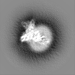
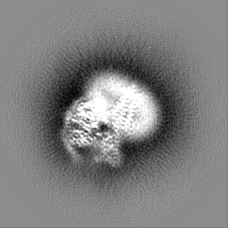
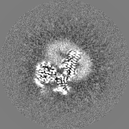
| 画像 |  emd_33503.png emd_33503.png | 43.9 KB | ||
| その他 | emd_33503_half_map_1.map.gzemd_33503_half_map_2.map.gz | 49.5 MB 49.5 MB | ||
| アーカイブディレクトリ |  http://ftp.pdbj.org/pub/emdb/structures/EMD-33503ftp://ftp.pdbj.org/pub/emdb/structures/EMD-33503 http://ftp.pdbj.org/pub/emdb/structures/EMD-33503ftp://ftp.pdbj.org/pub/emdb/structures/EMD-33503 | HTTPS FTP |
-関連構造データ

-リンク
| EMDBのページ | EMDB (EBI/PDBe) / EMDataResource |
|---|---|
| 「今月の分子」の関連する項目 |


-マップ
| ファイル | ダウンロード / ファイル: emd_33503.map.gz / 形式: CCP4 / 大きさ: 64 MB / タイプ: IMAGE STORED AS FLOATING POINT NUMBER (4 BYTES) | ||||||||||||||||||||
|---|---|---|---|---|---|---|---|---|---|---|---|---|---|---|---|---|---|---|---|---|---|
| ボクセルのサイズ | X=Y=Z: 1.045 Å | ||||||||||||||||||||
| 密度 |
| ||||||||||||||||||||
| 対称性 | 空間群: 1 | ||||||||||||||||||||
| 詳細 | EMDB XML:
|
-添付データ
-ハーフマップ: #2
| ファイル | emd_33503_half_map_1.map | ||||||||||||
|---|---|---|---|---|---|---|---|---|---|---|---|---|---|
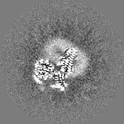
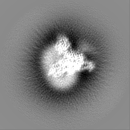
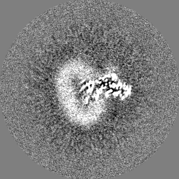
| 投影像・断面図 |
| ||||||||||||
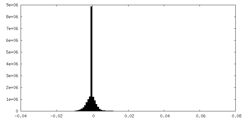
| 密度ヒストグラム |
 Z
Z Y
Y X
X


-ハーフマップ: #1
| ファイル | emd_33503_half_map_2.map | ||||||||||||
|---|---|---|---|---|---|---|---|---|---|---|---|---|---|
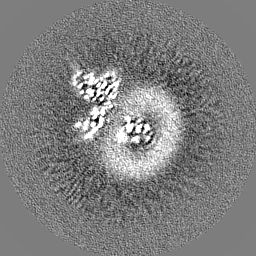
| 投影像・断面図 |
| ||||||||||||
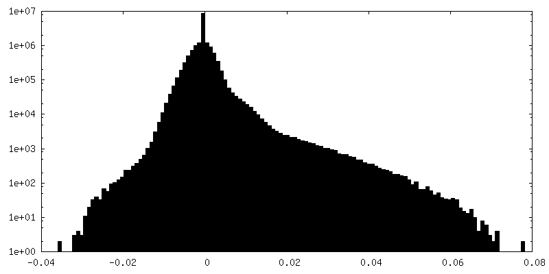
| 密度ヒストグラム |


- 試料の構成要素
-全体 : The purinergic receptor P2Y1R in complex with 2MeSADP, G11 and scfv16
| 全体 | 名称: The purinergic receptor P2Y1R in complex with 2MeSADP, G11 and scfv16 |
|---|---|
| 要素 |
|
-超分子 #1: The purinergic receptor P2Y1R in complex with 2MeSADP, G11 and scfv16
| 超分子 | 名称: The purinergic receptor P2Y1R in complex with 2MeSADP, G11 and scfv16 タイプ: complex / ID: 1 / 親要素: 0 / 含まれる分子: #2-#5 / 詳細: scf16 was recombinantly expressed in sf9 cells |
|---|---|
| 由来(天然) | 生物種: Homo sapiens (ヒト) |
| 分子量 | 理論値: 10 kDa/nm |
-分子 #1: P2Y purinoceptor 1
| 分子 | 名称: P2Y purinoceptor 1 / タイプ: protein_or_peptide / ID: 1 / コピー数: 1 / 光学異性体: LEVO |
|---|---|
| 由来(天然) | 生物種: Homo sapiens (ヒト) |
| 分子量 | 理論値: 46.296887 KDa |
| 組換発現 | 生物種:   Spodoptera frugiperda (ツマジロクサヨトウ) Spodoptera frugiperda (ツマジロクサヨトウ) |
| 配列 | 文字列: GAPTEVLWPA VPNGTDAAFL AGPGSSWGNS TVASTAAVSS SFKCALTKTG FQFYYLPAVY ILVFIIGFLG NSVAIWMFVF HMKPWSGIS VYMFNLALAD FLYVLTLPAL IFYYFNKTDW IFGDAMCKLQ RFIFHVNLYG SILFLTCISA HRYSGVVYPL K SLGRLKKK ...文字列: GAPTEVLWPA VPNGTDAAFL AGPGSSWGNS TVASTAAVSS SFKCALTKTG FQFYYLPAVY ILVFIIGFLG NSVAIWMFVF HMKPWSGIS VYMFNLALAD FLYVLTLPAL IFYYFNKTDW IFGDAMCKLQ RFIFHVNLYG SILFLTCISA HRYSGVVYPL K SLGRLKKK NAICISVLVW LIVVVAISPI LFYSGTGVRK NKTITCYDTT SDEYLRSYFI YSMCTTVAMF CVPLVLILGC YG LIVRALI YKDLDNSPLR RKSIYLVIIV LTVFAVSYIP FHVMKTMNLR ARLDFQTPAM CAFNDRVYAT YQVTRGLASL NSC VDPILY FLAGDTFRRR LSRATRKASR RSEANLQSKS EDMTLNILPE FKQNGDTSLE FLEVLFQGPG SWSHPQFEKG SGAG ASAGS WSHPQFEK UniProtKB: P2Y purinoceptor 1 |
-分子 #2: Guanine nucleotide-binding protein G(11) subunit alpha
| 分子 | 名称: Guanine nucleotide-binding protein G(11) subunit alpha タイプ: protein_or_peptide / ID: 2 / コピー数: 1 / 光学異性体: LEVO |
|---|---|
| 由来(天然) | 生物種: Homo sapiens (ヒト) |
| 分子量 | 理論値: 41.385281 KDa |
| 組換発現 | 生物種: Spodoptera frugiperda (ツマジロクサヨトウ) |
| 配列 | 文字列: MGCTLSAEDK AAVERSKMID RNLRRDKRDA RRELKLLLLG TGESGKSTFI KQMRIIHGAG YSEEDKRGFT KLVYQNIFTA MQAMIRAME TLKILYKYEQ NKANALLIRE VDVEKVTTFE HQYVSAIKTL WEDPGIQECY DRRREYQLSD SAKYYLTDVD R IATLGYLP ...文字列: MGCTLSAEDK AAVERSKMID RNLRRDKRDA RRELKLLLLG TGESGKSTFI KQMRIIHGAG YSEEDKRGFT KLVYQNIFTA MQAMIRAME TLKILYKYEQ NKANALLIRE VDVEKVTTFE HQYVSAIKTL WEDPGIQECY DRRREYQLSD SAKYYLTDVD R IATLGYLP TQQDVLRVRV PTTGIIEYPF DLENIIFRMV DVGGQRSERR KWIHCFENVT SIMFLVALSE YDQVLVESDN EN RMEESKA LFRTIITYPW FQNSSVILFL NKKDLLEDKI LYSHLVDYFP EFDGPQRDAQ AAREFILKMF VDLNPDSDKI IYS HFTCAT DTENIRFVFA AVKDTILQLN LKEYNLV |
-分子 #3: Guanine nucleotide-binding protein G(I)/G(S)/G(T) subunit beta-1
| 分子 | 名称: Guanine nucleotide-binding protein G(I)/G(S)/G(T) subunit beta-1 タイプ: protein_or_peptide / ID: 3 / コピー数: 1 / 光学異性体: LEVO |
|---|---|
| 由来(天然) | 生物種: Homo sapiens (ヒト) |
| 分子量 | 理論値: 38.245805 KDa |
| 組換発現 | 生物種: Spodoptera frugiperda (ツマジロクサヨトウ) |
| 配列 | 文字列: MHHHHHHSEL DQLRQEAEQL KNQIRDARKA CADATLSQIT NNIDPVGRIQ MRTRRTLRGH LAKIYAMHWG TDSRLLVSAS QDGKLIIWD SYTTNKVHAI PLRSSWVMTC AYAPSGNYVA CGGLDNICSI YNLKTREGNV RVSRELAGHT GYLSCCRFLD D NQIVTSSG ...文字列: MHHHHHHSEL DQLRQEAEQL KNQIRDARKA CADATLSQIT NNIDPVGRIQ MRTRRTLRGH LAKIYAMHWG TDSRLLVSAS QDGKLIIWD SYTTNKVHAI PLRSSWVMTC AYAPSGNYVA CGGLDNICSI YNLKTREGNV RVSRELAGHT GYLSCCRFLD D NQIVTSSG DTTCALWDIE TGQQTTTFTG HTGDVMSLSL APDTRLFVSG ACDASAKLWD VREGMCRQTF TGHESDINAI CF FPNGNAF ATGSDDATCR LFDLRADQEL MTYSHDNIIC GITSVSFSKS GRLLLAGYDD FNCNVWDALK ADRAGVLAGH DNR VSCLGV TDDGMAVATG SWDSFLKIWN UniProtKB: Guanine nucleotide-binding protein G(I)/G(S)/G(T) subunit beta-1 |
-分子 #4: Guanine nucleotide-binding protein G(I)/G(S)/G(O) subunit gamma-2
| 分子 | 名称: Guanine nucleotide-binding protein G(I)/G(S)/G(O) subunit gamma-2 タイプ: protein_or_peptide / ID: 4 / コピー数: 1 / 光学異性体: LEVO |
|---|---|
| 由来(天然) | 生物種: Homo sapiens (ヒト) |
| 分子量 | 理論値: 7.861143 KDa |
| 組換発現 | 生物種: Spodoptera frugiperda (ツマジロクサヨトウ) |
| 配列 | 文字列: MASNNTASIA QARKLVEQLK MEANIDRIKV SKAAADLMAY CEAHAKEDPL LTPVPASENP FREKKFFCAI L UniProtKB: Guanine nucleotide-binding protein G(I)/G(S)/G(O) subunit gamma-2 |
-分子 #5: scfv16
| 分子 | 名称: scfv16 / タイプ: protein_or_peptide / ID: 5 / コピー数: 1 / 光学異性体: LEVO |
|---|---|
| 由来(天然) | 生物種: Homo sapiens (ヒト) |
| 分子量 | 理論値: 26.337307 KDa |
| 組換発現 | 生物種: Spodoptera frugiperda (ツマジロクサヨトウ) |
| 配列 | 文字列: DVQLVESGGG LVQPGGSRKL SCSASGFAFS SFGMHWVRQA PEKGLEWVAY ISSGSGTIYY ADTVKGRFTI SRDDPKNTLF LQMTSLRSE DTAMYYCVRS IYYYGSSPFD FWGQGTTLTV SSGGGGSGGG GSGGGGSDIV MTQATSSVPV TPGESVSISC R SSKSLLHS ...文字列: DVQLVESGGG LVQPGGSRKL SCSASGFAFS SFGMHWVRQA PEKGLEWVAY ISSGSGTIYY ADTVKGRFTI SRDDPKNTLF LQMTSLRSE DTAMYYCVRS IYYYGSSPFD FWGQGTTLTV SSGGGGSGGG GSGGGGSDIV MTQATSSVPV TPGESVSISC R SSKSLLHS NGNTYLYWFL QRPGQSPQLL IYRMSNLASG VPDRFSGSGS GTAFTLTISR LEAEDVGVYY CMQHLEYPLT FG AGTKLEL |
-分子 #6: 2-(methylsulfanyl)adenosine 5'-(trihydrogen diphosphate)
| 分子 | 名称: 2-(methylsulfanyl)adenosine 5'-(trihydrogen diphosphate) タイプ: ligand / ID: 6 / コピー数: 1 / 式: 6AD |
|---|---|
| 分子量 | 理論値: 473.293 Da |
| Chemical component information | 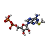 ChemComp-6AD: |
-実験情報
-構造解析
| 手法 | クライオ電子顕微鏡法 |
|---|---|
 解析 解析 | 単粒子再構成法 |
| 試料の集合状態 | particle |
-試料調製
| 濃度 | 2.0 mg/mL |
|---|---|
| 緩衝液 | pH: 7.5 / 構成要素 - 濃度: 2.0 mg/ml / 構成要素 - 式: 150mM / 構成要素 - 名称: sodium chloride塩化ナトリウム |
| グリッド | モデル: C-flat-1.2/1.3 / 材質: GOLD / メッシュ: 400 / 前処理 - タイプ: GLOW DISCHARGE |
| 凍結 | 凍結剤: ETHANE / チャンバー内湿度: 100 % / チャンバー内温度: 298 K / 装置: FEI VITROBOT MARK IV / 詳細: blot for 1s. |
| 詳細 | This sample was monodisperse |
- 電子顕微鏡法
| 顕微鏡 | FEI TITAN KRIOS |
|---|---|
| 電子線 | 加速電圧: 300 kV / 電子線源: FIELD EMISSION GUN |
| 電子光学系 | 照射モード: FLOOD BEAM / 撮影モード: BRIGHT FIELDBright-field microscopy / Cs: 2.7 mm / 最大 デフォーカス(公称値): 1.5 µm / 最小 デフォーカス(公称値): 0.8 µm |
| 撮影 | フィルム・検出器のモデル: GATAN K3 BIOQUANTUM (6k x 4k) 撮影したグリッド数: 2 / 実像数: 9856 / 平均露光時間: 2.0 sec. / 平均電子線量: 60.0 e/Å2 / 詳細: Images were collected in movie-mode |
| 実験機器 |  モデル: Titan Krios / 画像提供: FEI Company |
-画像解析
| 粒子像選択 | 選択した数: 6184568 |
|---|---|
| 初期モデル | モデルのタイプ: PDB ENTRY PDBモデル - PDB ID: 詳細: 6OIJ for G protein |
| 初期 角度割当 | タイプ: MAXIMUM LIKELIHOOD |
| 最終 3次元分類 | ソフトウェア - 名称: RELION (ver. 3.0) |
| 最終 角度割当 | タイプ: NOT APPLICABLE |
| 最終 再構成 | 想定した対称性 - 点群: C1 (非対称) / 解像度のタイプ: BY AUTHOR / 解像度: 2.9 Å / 解像度の算出法: FSC 0.143 CUT-OFF / 使用した粒子像数: 659399 |
| 詳細 | The selected images were 20eV using GIF-Quantum LS Imaging energy filter |

