 Movie
Movie Controller

[English] 日本語
 Yorodumi
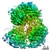
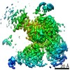
- EMDB-30638: cryo-EM structure of the DEAH-box helicase Prp2 and coactivator Spp2 -
+ Open data

- Basic information
| Entry | Database: EMDB / ID: EMD-30638 | |||||||||
|---|---|---|---|---|---|---|---|---|---|---|
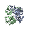
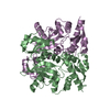
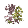
| Title | cryo-EM structure of the DEAH-box helicase Prp2 and coactivator Spp2 | |||||||||
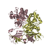
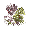
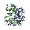
 Map data Map data | EM map of ADP bound Prp2-Spp2 complex at 2.88 angstrom | |||||||||
 Sample Sample |
| |||||||||
 Keywords Keywords |  spliceosome / RNA splicing / Bact complex / DEAH-box ATPase/helicase / Prp2 / Spp2 / SPLICING spliceosome / RNA splicing / Bact complex / DEAH-box ATPase/helicase / Prp2 / Spp2 / SPLICING | |||||||||
| Function / homology |  Function and homology information Function and homology informationsnoRNA splicing / generation of catalytic spliceosome for first transesterification step / ATP-dependent activity, acting on RNA / U2-type catalytic step 1 spliceosome / ATPase activator activity / catalytic step 2 spliceosome / helicase activity / spliceosomal complex / mRNA splicing, via spliceosome / nucleic acid binding ...snoRNA splicing / generation of catalytic spliceosome for first transesterification step / ATP-dependent activity, acting on RNA / U2-type catalytic step 1 spliceosome / ATPase activator activity / catalytic step 2 spliceosome / helicase activity / spliceosomal complex / mRNA splicing, via spliceosome / nucleic acid binding / RNA helicase activity / RNA helicase / ATP hydrolysis activity / RNA binding / ATP binding / nucleus / cytoplasmSimilarity search - Function | |||||||||
| Biological species |  Saccharomyces cerevisiae (brewer's yeast) Saccharomyces cerevisiae (brewer's yeast) | |||||||||
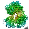
| Method | single particle reconstruction / cryo EM / Resolution: 3.15 Å | |||||||||
 Authors Authors | Bai R / Wan R | |||||||||
| Funding support |  China, 1 items China, 1 items
| |||||||||
 Citation Citation | Journal: Science / Year: 2021 Title: Mechanism of spliceosome remodeling by the ATPase/helicase Prp2 and its coactivator Spp2. Authors: Rui Bai / Ruixue Wan / Chuangye Yan / Qi Jia / Jianlin Lei / Yigong Shi / Abstract: Spliceosome remodeling, executed by conserved adenosine triphosphatase (ATPase)/helicases including Prp2, enables precursor messenger RNA (pre-mRNA) splicing. However, the structural basis for the ...Spliceosome remodeling, executed by conserved adenosine triphosphatase (ATPase)/helicases including Prp2, enables precursor messenger RNA (pre-mRNA) splicing. However, the structural basis for the function of the ATPase/helicases remains poorly understood. Here, we report atomic structures of Prp2 in isolation, Prp2 complexed with its coactivator Spp2, and Prp2-loaded activated spliceosome and the results of structure-guided biochemical analysis. Prp2 weakly associates with the spliceosome and cannot function without Spp2, which stably associates with Prp2 and anchors on the spliceosome, thus tethering Prp2 to the activated spliceosome and allowing Prp2 to function. Pre-mRNA is loaded into a featured channel between the N and C halves of Prp2, where Leu from the N half and Arg from the C half prevent backward sliding of pre-mRNA toward its 5'-end. Adenosine 5'-triphosphate binding and hydrolysis trigger interdomain movement in Prp2, which drives unidirectional stepwise translocation of pre-mRNA toward its 3'-end. These conserved mechanisms explain the coupling of spliceosome remodeling to pre-mRNA splicing. | |||||||||
| History |
|
- Structure visualization
| Movie |
Movie viewer |
|---|---|
| Structure viewer | EM map: SurfViewMolmilJmol/JSmol |
| Supplemental images |

- Downloads & links
-EMDB archive
| Map data | emd_30638.map.gz | 96.5 MB | EMDB map data format | |
|---|---|---|---|---|
| Header (meta data) | emd-30638-v30.xmlemd-30638.xml | 12.6 KB 12.6 KB | Display Display | EMDB header |
| Images | 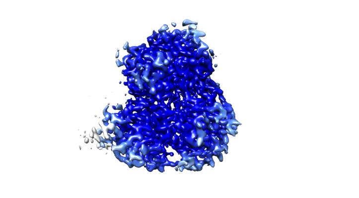 emd_30638.png emd_30638.png | 155.5 KB | ||
| Filedesc metadata | emd-30638.cif.gz | 6.1 KB | ||
| Archive directory |  http://ftp.pdbj.org/pub/emdb/structures/EMD-30638ftp://ftp.pdbj.org/pub/emdb/structures/EMD-30638 http://ftp.pdbj.org/pub/emdb/structures/EMD-30638ftp://ftp.pdbj.org/pub/emdb/structures/EMD-30638 | HTTPS FTP |
-Related structure data
| Related structure data | 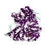 7dcpMC 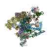 7dcoC 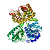 7dcqC 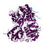 7dcrC 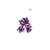 7dd3C M: atomic model generated by this map C: citing same article ( |
|---|---|
| Similar structure data |


-Links
| EMDB pages | EMDB (EBI/PDBe) / EMDataResource |
|---|---|
| Related items in Molecule of the Month |

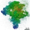
-Map
| File | Download / File: emd_30638.map.gz / Format: CCP4 / Size: 103 MB / Type: IMAGE STORED AS FLOATING POINT NUMBER (4 BYTES) | ||||||||||||||||||||||||||||||||||||||||||||||||||||||||||||
|---|---|---|---|---|---|---|---|---|---|---|---|---|---|---|---|---|---|---|---|---|---|---|---|---|---|---|---|---|---|---|---|---|---|---|---|---|---|---|---|---|---|---|---|---|---|---|---|---|---|---|---|---|---|---|---|---|---|---|---|---|---|
| Annotation | EM map of ADP bound Prp2-Spp2 complex at 2.88 angstrom | ||||||||||||||||||||||||||||||||||||||||||||||||||||||||||||
| Voxel size | X=Y=Z: 0.6625 Å | ||||||||||||||||||||||||||||||||||||||||||||||||||||||||||||
| Density |
| ||||||||||||||||||||||||||||||||||||||||||||||||||||||||||||
| Symmetry | Space group: 1 | ||||||||||||||||||||||||||||||||||||||||||||||||||||||||||||
| Details | EMDB XML:
CCP4 map header:
| ||||||||||||||||||||||||||||||||||||||||||||||||||||||||||||
-Supplemental data
- Sample components
-Entire : Complex of ATPase/helicase Prp2 and its coactivator Spp2
| Entire | Name: Complex of ATPase/helicase Prp2 and its coactivator Spp2 |
|---|---|
| Components |
|
-Supramolecule #1: Complex of ATPase/helicase Prp2 and its coactivator Spp2
| Supramolecule | Name: Complex of ATPase/helicase Prp2 and its coactivator Spp2 type: complex / ID: 1 / Parent: 0 / Macromolecule list: #1-#2 |
|---|---|
| Source (natural) | Organism: Saccharomyces cerevisiae (brewer's yeast) |
| Molecular weight | Theoretical: 119 kDa/nm |
-Macromolecule #1: PRP2 isoform 1
| Macromolecule | Name: PRP2 isoform 1 / type: protein_or_peptide / ID: 1 / Number of copies: 1 / Enantiomer: LEVO |
|---|---|
| Source (natural) | Organism: Saccharomyces cerevisiae (brewer's yeast) |
| Molecular weight | Theoretical: 97.663961 KDa |
| Recombinant expression | Organism:  Escherichia coli BL21(DE3) (bacteria) Escherichia coli BL21(DE3) (bacteria) |
| Sequence | String: RVKRTYEVTR QNDNAVRIEP SSLGEEEDKE AKDKNSALQL KRSRYDPNKV FSNTNQGPEK NNLKGEQLGS QKKSSKYDEK ITSNNELTT KKGLLGDSEN ETKYASSNSK FNVEVTHKIK NAKEIDKINR QRMWEEQQLR NAMAGQSDHP DDITLEGSDK Y DYVFDTDA ...String: RVKRTYEVTR QNDNAVRIEP SSLGEEEDKE AKDKNSALQL KRSRYDPNKV FSNTNQGPEK NNLKGEQLGS QKKSSKYDEK ITSNNELTT KKGLLGDSEN ETKYASSNSK FNVEVTHKIK NAKEIDKINR QRMWEEQQLR NAMAGQSDHP DDITLEGSDK Y DYVFDTDA MIDYTNEEDD LLPEEKLQYE ARLAQALETE EKRILTIQEA RKLLPVHQYK DELLQEIKKN QVLIIMGETG SG KTTQLPQ YLVEDGFTDQ GKLQIAITQP RRVAATSVAA RVADEMNVVL GKEVGYQIRF EDKTTPNKTV LKYMTDGMLL REF LTDSKL SKYSCIMIDE AHERTLATDI LIGLLKDILP QRPTLKLLIS SATMNAKKFS EFFDNCPIFN VPGRRYPVDI HYTL QPEAN YIHAAITTIF QIHTTQSLPG DILVFLTGQE EIERTKTKLE EIMSKLGSRT KQMIITPIYA NLPQEQQLKI FQPTP ENCR KVVLATNIAE TSLTIDGIRY VIDPGFVKEN SYVPSTGMTQ LLTVPCSRAS VDQRAGRAGR VGPGKCFRIF TKWSYL HEL ELMPKPEITR TNLSNTVLLL LSLGVTDLIK FPLMDKPSIP TLRKSLENLY ILGALNSKGT ITRLGKMMCE FPCEPEF AK VLYTAATHEQ CQGVLEECLT IVSMLHETPS LFIGQKRDAA ASVLSEVESD HILYLEIFNQ WRNSKFSRSW CQDHKIQF K TMLRVRNIRN QLFRCSEKVG LVEKNDQARM KIGNIAGYIN ARITRCFISG FPMNIVQLGP TGYQTMGRSS GGLNVSVHP TSILFVNHKE KAQRPSKYVL YQQLMLTSKE FIRDCLVIPK EEWLIDMVPQ IFKDLI UniProtKB: PRP2 isoform 1 |
-Macromolecule #2: Pre-mRNA-splicing factor SPP2
| Macromolecule | Name: Pre-mRNA-splicing factor SPP2 / type: protein_or_peptide / ID: 2 / Number of copies: 1 / Enantiomer: LEVO |
|---|---|
| Source (natural) | Organism: Saccharomyces cerevisiae (brewer's yeast) |
| Molecular weight | Theoretical: 20.685377 KDa |
| Recombinant expression | Organism: Escherichia coli BL21(DE3) (bacteria) |
| Sequence | String: MSKFSLKLGS KTLKKNISKK TKKKNSLQKA NLFDWDDAET ASLSHKPQSK IKIQSIDKFD LDEESSSKKK LVIKLSENAD TKKNDAPLV EYVTEKEYNE VPVEEFGDAL LRGMGWESDS EQDSKGDKTQ SRNKDVSNVS QIHPDGLGIG AKLNKAINVE E ASFMPVVK IDKITGTKVD DDKKNKS UniProtKB: Pre-mRNA-splicing factor SPP2 |
-Macromolecule #3: ADENOSINE-5'-DIPHOSPHATE
| Macromolecule | Name: ADENOSINE-5'-DIPHOSPHATE / type: ligand / ID: 3 / Number of copies: 1 / Formula: ADP |
|---|---|
| Molecular weight | Theoretical: 427.201 Da |
| Chemical component information |  ChemComp-ADP: |
-Macromolecule #4: MAGNESIUM ION
| Macromolecule | Name: MAGNESIUM ION / type: ligand / ID: 4 / Number of copies: 1 / Formula: MG |
|---|---|
| Molecular weight | Theoretical: 24.305 Da |
-Experimental details
-Structure determination
| Method | cryo EM |
|---|---|
 Processing Processing | single particle reconstruction |
| Aggregation state | particle |
-Sample preparation
| Buffer | pH: 7.9 |
|---|---|
| Grid | Model: Quantifoil R1.2/1.3 / Material: GOLD / Mesh: 300 / Support film - Material: GRAPHENE / Support film - topology: CONTINUOUS / Pretreatment - Type: GLOW DISCHARGE / Pretreatment - Time: 10 sec. |
| Vitrification | Cryogen name: ETHANE / Chamber humidity: 100 % |
- Electron microscopy
| Microscope | FEI TITAN |
|---|---|
| Electron beam | Acceleration voltage: 300 kV / Electron source: FIELD EMISSION GUN |
| Electron optics | Illumination mode: FLOOD BEAM / Imaging mode: BRIGHT FIELDBright-field microscopy |
| Specialist optics | Energy filter - Slit width: 20 eV |
| Image recording | Film or detector model: GATAN K3 (6k x 4k) / Average electron dose: 50.0 e/Å2 |
-Image processing
| Startup model | Type of model: EMDB MAP |
|---|---|
| Initial angle assignment | Type: MAXIMUM LIKELIHOOD |
| Final angle assignment | Type: MAXIMUM LIKELIHOOD |
| Final reconstruction | Applied symmetry - Point group: C1 (asymmetric) / Resolution.type: BY AUTHOR / Resolution: 3.15 Å / Resolution method: FSC 0.143 CUT-OFF / Number images used: 130302 |
