 Movie
Movie Controller

[English] 日本語
 Yorodumi
- EMDB-2973: electron crystallographic structure of lens Aquaporin-0 (AQP0) (l... -
+ Open data

- Basic information
| Entry | Database: EMDB / ID: EMD-2973 | |||||||||
|---|---|---|---|---|---|---|---|---|---|---|
| Title | electron crystallographic structure of lens Aquaporin-0 (AQP0) (lens MIP) at 1.9A resolution, in a closed pore state | |||||||||
 Map data Map data | complete unit cell. AQP0 2Fo-Fc map | |||||||||
 Sample Sample |
| |||||||||
 Keywords Keywords | Aquaporin-0 / electron crystallography / membrane channel | |||||||||
| Function / homology |  Function and homology information Function and homology informationgap junction-mediated intercellular transport / water transport / water channel activity / structural constituent of eye lens / gap junction / lens development in camera-type eye / positive regulation of cell adhesion / visual perception / protein homotetramerization / calmodulin binding ...gap junction-mediated intercellular transport / water transport / water channel activity / structural constituent of eye lens / gap junction / lens development in camera-type eye / positive regulation of cell adhesion / visual perception / protein homotetramerization / calmodulin binding / apical plasma membrane / endoplasmic reticulum / plasma membrane Similarity search - Function | |||||||||
| Biological species |  | |||||||||
| Method | electron crystallography / cryo EM / Resolution: 1.9 Å | |||||||||
 Authors Authors | Gonen T / Cheng Y / Sliz P / Hiroaki Y / Fujiyoshi Y / Harrison SC / Walz T | |||||||||
 Citation Citation | Journal: Nature / Year: 2005 Title: Lipid-protein interactions in double-layered two-dimensional AQP0 crystals. Authors: Tamir Gonen / Yifan Cheng / Piotr Sliz / Yoko Hiroaki / Yoshinori Fujiyoshi / Stephen C Harrison / Thomas Walz /  Abstract: Lens-specific aquaporin-0 (AQP0) functions as a specific water pore and forms the thin junctions between fibre cells. Here we describe a 1.9 A resolution structure of junctional AQP0, determined by ...Lens-specific aquaporin-0 (AQP0) functions as a specific water pore and forms the thin junctions between fibre cells. Here we describe a 1.9 A resolution structure of junctional AQP0, determined by electron crystallography of double-layered two-dimensional crystals. Comparison of junctional and non-junctional AQP0 structures shows that junction formation depends on a conformational switch in an extracellular loop, which may result from cleavage of the cytoplasmic amino and carboxy termini. In the centre of the water pathway, the closed pore in junctional AQP0 retains only three water molecules, which are too widely spaced to form hydrogen bonds with each other. Packing interactions between AQP0 tetramers in the crystalline array are mediated by lipid molecules, which assume preferred conformations. We were therefore able to build an atomic model for the lipid bilayer surrounding the AQP0 tetramers, and we describe lipid-protein interactions. | |||||||||
| History |
|
- Structure visualization
| Movie |
Movie viewer |
|---|---|
| Structure viewer | EM map: SurfViewMolmilJmol/JSmol |
| Supplemental images |

- Downloads & links
-EMDB archive
| Map data | emd_2973.map.gz | 25.6 MB | EMDB map data format | |
|---|---|---|---|---|
| Header (meta data) | emd-2973-v30.xmlemd-2973.xml | 12.7 KB 12.7 KB | Display Display | EMDB header |
| Images | 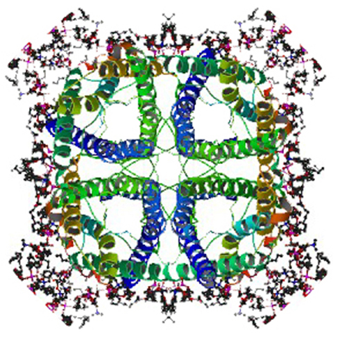 emd_2973.jpg emd_2973.jpg | 280.8 KB | ||
| Masks | emd_2973_msk_1.map | 9.1 MB | Mask map | |
| Filedesc structureFactors | emd_2973_sf.cif.gz | 157.7 KB | ||
| Archive directory |  http://ftp.pdbj.org/pub/emdb/structures/EMD-2973ftp://ftp.pdbj.org/pub/emdb/structures/EMD-2973 http://ftp.pdbj.org/pub/emdb/structures/EMD-2973ftp://ftp.pdbj.org/pub/emdb/structures/EMD-2973 | HTTPS FTP |
-Validation report
| Summary document | emd_2973_validation.pdf.gz | 309.1 KB | Display | EMDB validaton report |
|---|---|---|---|---|
| Full document | emd_2973_full_validation.pdf.gz | 308.3 KB | Display | |
| Data in XML | emd_2973_validation.xml.gz | 4.5 KB | Display | |
| Arichive directory | https://ftp.pdbj.org/pub/emdb/validation_reports/EMD-2973ftp://ftp.pdbj.org/pub/emdb/validation_reports/EMD-2973 | HTTPS FTP |
-Related structure data

-Links
| EMDB pages | EMDB (EBI/PDBe) / EMDataResource |
|---|---|
| Related items in Molecule of the Month |

-Map
| File | Download / File: emd_2973.map.gz / Format: CCP4 / Size: 27.8 MB / Type: IMAGE STORED AS FLOATING POINT NUMBER (4 BYTES) | ||||||||||||||||||||||||||||||||||||||||||||||||||||||||||||||||||||
|---|---|---|---|---|---|---|---|---|---|---|---|---|---|---|---|---|---|---|---|---|---|---|---|---|---|---|---|---|---|---|---|---|---|---|---|---|---|---|---|---|---|---|---|---|---|---|---|---|---|---|---|---|---|---|---|---|---|---|---|---|---|---|---|---|---|---|---|---|---|
| Annotation | complete unit cell. AQP0 2Fo-Fc map | ||||||||||||||||||||||||||||||||||||||||||||||||||||||||||||||||||||
| Voxel size | X: 0.4618 Å / Y: 0.4618 Å / Z: 0.4444 Å | ||||||||||||||||||||||||||||||||||||||||||||||||||||||||||||||||||||
| Density |
| ||||||||||||||||||||||||||||||||||||||||||||||||||||||||||||||||||||
| Symmetry | Space group: 89 | ||||||||||||||||||||||||||||||||||||||||||||||||||||||||||||||||||||
| Details | EMDB XML:
CCP4 map header:
| ||||||||||||||||||||||||||||||||||||||||||||||||||||||||||||||||||||
-Supplemental data
-Segmentation: 2Fo-Fc map of AQP0 determined by electron crystallography...
| Annotation | 2Fo-Fc map of AQP0 determined by electron crystallography - asymmetric unit only | ||||||||||||
|---|---|---|---|---|---|---|---|---|---|---|---|---|---|
| File | emd_2973_msk_1.map | ||||||||||||
| Projections & Slices |
| ||||||||||||
| Density Histograms |
 Z
Z Y
Y X
X


- Sample components
-Entire : Mammalian Aquaporin-0 (Sheep)
| Entire | Name: Mammalian Aquaporin-0 (Sheep) |
|---|---|
| Components |
|
-Supramolecule #1000: Mammalian Aquaporin-0 (Sheep)
| Supramolecule | Name: Mammalian Aquaporin-0 (Sheep) / type: sample / ID: 1000 / Oligomeric state: two tetramers / Number unique components: 1 |
|---|---|
| Molecular weight | Experimental: 120 KDa / Theoretical: 120 KDa / Method: SDS PAGE |
-Macromolecule #1: Aquaporin-0
| Macromolecule | Name: Aquaporin-0 / type: protein_or_peptide / ID: 1 / Name.synonym: Lens MIP, MP26 / Number of copies: 8 / Oligomeric state: two tetramers / Recombinant expression: No / Database: NCBI |
|---|---|
| Source (natural) | Organism: |
| Molecular weight | Experimental: 120 KDa / Theoretical: 120 KDa |
-Experimental details
-Structure determination
| Method | cryo EM |
|---|---|
 Processing Processing | electron crystallography |
| Aggregation state | 2D array |
-Sample preparation
| Concentration | 5 mg/mL |
|---|---|
| Buffer | pH: 8 / Details: 20mM Tris pH 8, 150mM NaCl, 25mM MgCl2 |
| Grid | Details: Mo hexagonal grids |
| Vitrification | Cryogen name: HELIUM / Chamber temperature: 4 K / Instrument: OTHER |
| Details | dialysis into 2d sheets |
| Crystal formation | Details: dialysis into 2d sheets |
- Electron microscopy
| Microscope | JEOL 3200FSC |
|---|---|
| Temperature | Min: 4 K / Max: 30 K / Average: 8 K |
| Specialist optics | Energy filter - Name: omega filter JEOL |
| Date | Oct 9, 2004 |
| Image recording | Category: CCD / Film or detector model: TVIPS TEMCAM-F416 (4k x 4k) / Number real images: 286 / Average electron dose: 20 e/Å2 / Camera length: 1500 |
| Tilt angle min | 0 |
| Electron beam | Acceleration voltage: 300 kV / Electron source:  FIELD EMISSION GUN FIELD EMISSION GUN |
| Electron optics | Illumination mode: FLOOD BEAM / Imaging mode: DIFFRACTION / Cs: 1.2 mm |
| Sample stage | Specimen holder: Helium Cooled / Specimen holder model: JEOL 3200FSC CRYOHOLDER / Tilt angle max: 70 / Tilt series - Axis1 - Min angle: 0 ° / Tilt series - Axis1 - Max angle: 70 ° |
-Image processing
| Details | Data was processed in XDP. Model building in O and refinement in CNS. |
|---|---|
| Final reconstruction | Resolution.type: BY AUTHOR / Resolution: 1.9 Å / Resolution method: DIFFRACTION PATTERN/LAYERLINES / Details: This submission corresponds to the PDB entry 2B6O. |
| Crystal parameters | Unit cell - A: 65.5 Å / Unit cell - B: 65.5 Å / Unit cell - C: 160 Å / Unit cell - γ: 90 ° / Unit cell - α: 90.0 ° / Unit cell - β: 90.0 ° / Plane group: P 4 2 2 |
