 Movie
Movie Controller


+ Open data

- Basic information
| Entry | Database: EMDB / ID: EMD-20842 | |||||||||
|---|---|---|---|---|---|---|---|---|---|---|
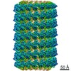
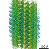
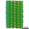
| Title | Endophilin B1 helical scaffold | |||||||||
 Map data Map data | Helical assembly of endophilin B1_N-BAR | |||||||||
 Sample Sample |
| |||||||||
 Keywords Keywords |  Membrane binding / amphipathic helix / BAR protein / SH3 domain / membrane trafficking / cell death / CYTOSOLIC PROTEIN Membrane binding / amphipathic helix / BAR protein / SH3 domain / membrane trafficking / cell death / CYTOSOLIC PROTEIN | |||||||||
| Function / homology |  Function and homology information Function and homology informationprotein localization to vacuolar membrane / positive regulation of membrane tubulation / autophagic cell death / positive regulation of autophagosome assembly / receptor catabolic process / membrane fission / membrane organization / positive regulation of protein targeting to mitochondrion / autophagosome membrane / regulation of macroautophagy ...protein localization to vacuolar membrane / positive regulation of membrane tubulation / autophagic cell death / positive regulation of autophagosome assembly / receptor catabolic process / membrane fission / membrane organization / positive regulation of protein targeting to mitochondrion / autophagosome membrane / regulation of macroautophagy / cellular response to glucose starvation / positive regulation of autophagy / cellular response to amino acid starvation / regulation of cytokinesis / positive regulation of protein-containing complex assembly / regulation of protein stability / autophagy / midbody / cytoplasmic vesicle / mitochondrial outer membrane / cadherin binding / Golgi membrane / lipid binding / apoptotic process / protein homodimerization activity / protein-containing complex / membrane / identical protein binding / cytosol / cytoplasmSimilarity search - Function | |||||||||
| Biological species |  Homo sapiens (human) Homo sapiens (human) | |||||||||
| Method | helical reconstruction / cryo EM / Resolution: 10.0 Å | |||||||||
 Authors Authors | Bhatt VS / Sundborger-Lunna AC | |||||||||
 Citation Citation | Journal: Structure / Year: 2021 Title: Amphipathic Motifs Regulate N-BAR Protein Endophilin B1 Auto-inhibition and Drive Membrane Remodeling. Authors: Veer S Bhatt / Robert Ashley / Anna Sundborger-Lunna /  Abstract: Membrane remodeling is a common theme in a variety of cellular processes. Here, we investigated membrane remodeling N-BAR protein endophilin B1, a critical player in diverse intracellular trafficking ...Membrane remodeling is a common theme in a variety of cellular processes. Here, we investigated membrane remodeling N-BAR protein endophilin B1, a critical player in diverse intracellular trafficking events, including mitochondrial and Golgi fission, and apoptosis. We find that endophilin B1 assembles into helical scaffolds on membranes, and that both membrane binding and assembly are driven by interactions between N-terminal helix H0 and the lipid bilayer. Furthermore, we find that endophilin B1 membrane remodeling is auto-inhibited and identify direct SH3 domain-H0 interactions as the underlying mechanism. Our results indicate that lipid composition plays a role in dictating endophilin B1 activity. Taken together, this study provides insight into a poorly understood N-BAR protein family member and highlights molecular mechanisms that may be general for the regulation of membrane remodeling. Our work suggests that interplay between membrane lipids and membrane interacting proteins facilitates spatial and temporal coordination of membrane remodeling. | |||||||||
| History |
|
- Structure visualization
| Movie |
Movie viewer |
|---|---|
| Structure viewer | EM map: SurfViewMolmilJmol/JSmol |
| Supplemental images |

- Downloads & links
-EMDB archive
| Map data | emd_20842.map.gz | 35.2 MB | EMDB map data format | |
|---|---|---|---|---|
| Header (meta data) | emd-20842-v30.xmlemd-20842.xml | 9.3 KB 9.3 KB | Display Display | EMDB header |
| Images |  emd_20842.png emd_20842.png | 55.3 KB | ||
| Filedesc metadata | emd-20842.cif.gz | 5 KB | ||
| Archive directory |  http://ftp.pdbj.org/pub/emdb/structures/EMD-20842ftp://ftp.pdbj.org/pub/emdb/structures/EMD-20842 http://ftp.pdbj.org/pub/emdb/structures/EMD-20842ftp://ftp.pdbj.org/pub/emdb/structures/EMD-20842 | HTTPS FTP |
-Related structure data
| Related structure data |  6upnMC 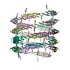 6up6C C: citing same article ( M: atomic model generated by this map |
|---|---|
| Similar structure data |


-Links
| EMDB pages | EMDB (EBI/PDBe) / EMDataResource |
|---|---|
| Related items in Molecule of the Month |

-Map
| File | Download / File: emd_20842.map.gz / Format: CCP4 / Size: 37.4 MB / Type: IMAGE STORED AS FLOATING POINT NUMBER (4 BYTES) | ||||||||||||||||||||||||||||||||||||||||||||||||||||||||||||
|---|---|---|---|---|---|---|---|---|---|---|---|---|---|---|---|---|---|---|---|---|---|---|---|---|---|---|---|---|---|---|---|---|---|---|---|---|---|---|---|---|---|---|---|---|---|---|---|---|---|---|---|---|---|---|---|---|---|---|---|---|---|
| Annotation | Helical assembly of endophilin B1_N-BAR | ||||||||||||||||||||||||||||||||||||||||||||||||||||||||||||
| Voxel size | X=Y=Z: 2.2942 Å | ||||||||||||||||||||||||||||||||||||||||||||||||||||||||||||
| Density |
| ||||||||||||||||||||||||||||||||||||||||||||||||||||||||||||
| Symmetry | Space group: 1 | ||||||||||||||||||||||||||||||||||||||||||||||||||||||||||||
| Details | EMDB XML:
CCP4 map header:
| ||||||||||||||||||||||||||||||||||||||||||||||||||||||||||||
-Supplemental data
- Sample components
-Entire : Endophilin B1 helical assembly
| Entire | Name: Endophilin B1 helical assembly |
|---|---|
| Components |
|
-Supramolecule #1: Endophilin B1 helical assembly
| Supramolecule | Name: Endophilin B1 helical assembly / type: complex / ID: 1 / Parent: 0 / Macromolecule list: all / Details: Endohilin B1 N-BAR domain |
|---|---|
| Source (natural) | Organism: Homo sapiens (human) |
-Macromolecule #1: Endophilin-B1
| Macromolecule | Name: Endophilin-B1 / type: protein_or_peptide / ID: 1 / Number of copies: 48 / Enantiomer: LEVO |
|---|---|
| Source (natural) | Organism: Homo sapiens (human) |
| Molecular weight | Theoretical: 40.843246 KDa |
| Recombinant expression | Organism:  Escherichia coli (E. coli) Escherichia coli (E. coli) |
| Sequence | String: MNIMDFNVKK LAADAGTFLS RAVQFTEEKL GQAEKTELDA HLENLLSKAE CTKIWTEKIM KQTEVLLQPN PNARIEEFVY EKLDRKAPS RINNPELLGQ YMIDAGTEFG PGTAYGNALI KCGETQKRIG TADRELIQTS ALNFLTPLRN FIEGDYKTIA K ERKLLQNK ...String: MNIMDFNVKK LAADAGTFLS RAVQFTEEKL GQAEKTELDA HLENLLSKAE CTKIWTEKIM KQTEVLLQPN PNARIEEFVY EKLDRKAPS RINNPELLGQ YMIDAGTEFG PGTAYGNALI KCGETQKRIG TADRELIQTS ALNFLTPLRN FIEGDYKTIA K ERKLLQNK RLDLDAAKTR LKKAKAAETR NSSEQELRIT QSEFDRQAEI TRLLLEGISS THAHHLRCLN DFVEAQMTYY AQ CYQYMLD LQKQLGSFPS NYLSNNNQTS VTPVPSVLPN AIGSSAMAST SGLVITSPSN LSDLKECSGS RKARVLYDYD AAN STELSL LADEVITVFS VVGMDSDWLM GERGNQKGKV PITYLELLN UniProtKB: Endophilin-B1 |
-Experimental details
-Structure determination
| Method | cryo EM |
|---|---|
 Processing Processing | helical reconstruction |
| Aggregation state | helical array |
-Sample preparation
| Buffer | pH: 8.1 |
|---|---|
| Vitrification | Cryogen name: ETHANE |
- Electron microscopy
| Microscope | FEI TITAN KRIOS |
|---|---|
| Electron beam | Acceleration voltage: 300 kV / Electron source: FIELD EMISSION GUN |
| Electron optics | Illumination mode: FLOOD BEAM / Imaging mode: BRIGHT FIELDBright-field microscopy / Cs: 2.7 mm |
| Image recording | Film or detector model: FEI FALCON III (4k x 4k) / Detector mode: OTHER / Average electron dose: 30.0 e/Å2 |
| Experimental equipment |  Model: Titan Krios / Image courtesy: FEI Company |
-Image processing
| Startup model | Type of model: OTHER / Details: Featureless cylinder |
|---|---|
| Final angle assignment | Type: NOT APPLICABLE |
| Final reconstruction | Applied symmetry - Helical parameters - Δz: 17.8 Å Applied symmetry - Helical parameters - Δ&Phi: 59.1 ° Applied symmetry - Helical parameters - Axial symmetry: C1 (asymmetric) Resolution.type: BY AUTHOR / Resolution: 10.0 Å / Resolution method: FSC 0.143 CUT-OFF / Number images used: 21197 |
