 ムービー
ムービー コントローラー


+ データを開く

- 基本情報
基本情報
| 登録情報 | データベース: EMDB / ID: EMD-20711 | |||||||||
|---|---|---|---|---|---|---|---|---|---|---|
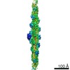
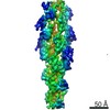
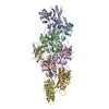
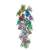
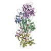
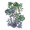
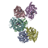
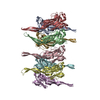
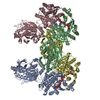
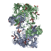
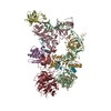
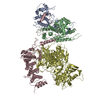
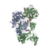
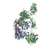
| タイトル | Cryo-EM structure of cofilactin from partially cofilin-decorated actin filaments. | |||||||||
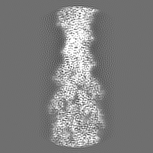
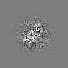
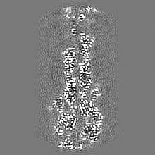
 マップデータ マップデータ | Helical reconstruction of cofilactin from a partially decorated sample. The map has been masked and sharpened with a b-factor of -215. | |||||||||
 試料 試料 |
| |||||||||
| 機能・相同性 |  機能・相同性情報 機能・相同性情報actin filament fragmentation / positive regulation of embryonic development / establishment of spindle localization / actin filament severing / regulation of dendritic spine morphogenesis / positive regulation by host of viral process / actin filament depolymerization / RHO GTPases Activate ROCKs / regulation of cell morphogenesis / cytoskeletal motor activator activity ...actin filament fragmentation / positive regulation of embryonic development / establishment of spindle localization / actin filament severing / regulation of dendritic spine morphogenesis / positive regulation by host of viral process / actin filament depolymerization / RHO GTPases Activate ROCKs / regulation of cell morphogenesis / cytoskeletal motor activator activity /  tropomyosin binding / mesenchyme migration / myosin heavy chain binding / troponin I binding / filamentous actin / actin filament bundle / lamellipodium membrane / skeletal muscle thin filament assembly / striated muscle thin filament / actin filament bundle assembly / Rho protein signal transduction / Sema3A PAK dependent Axon repulsion / mitotic cytokinesis / skeletal muscle myofibril / actin monomer binding / skeletal muscle fiber development / stress fiber / titin binding / cytoskeleton organization / EPHB-mediated forward signaling / actin filament polymerization / Gene and protein expression by JAK-STAT signaling after Interleukin-12 stimulation / filopodium / マイクロフィラメント / 加水分解酵素; 酸無水物に作用; 酸無水物に作用・細胞または細胞小器官の運動に関与 / response to virus / Regulation of actin dynamics for phagocytic cup formation / ruffle membrane / nuclear matrix / calcium-dependent protein binding / actin filament binding / マイクロフィラメント / Platelet degranulation / lamellipodium / cell body / 成長円錐 / actin cytoskeleton organization / vesicle / hydrolase activity / protein domain specific binding / focal adhesion / calcium ion binding / positive regulation of gene expression / negative regulation of apoptotic process / magnesium ion binding / extracellular space / extracellular exosome / ATP binding / 生体膜 / identical protein binding / 細胞核 / 細胞質基質 / 細胞質 tropomyosin binding / mesenchyme migration / myosin heavy chain binding / troponin I binding / filamentous actin / actin filament bundle / lamellipodium membrane / skeletal muscle thin filament assembly / striated muscle thin filament / actin filament bundle assembly / Rho protein signal transduction / Sema3A PAK dependent Axon repulsion / mitotic cytokinesis / skeletal muscle myofibril / actin monomer binding / skeletal muscle fiber development / stress fiber / titin binding / cytoskeleton organization / EPHB-mediated forward signaling / actin filament polymerization / Gene and protein expression by JAK-STAT signaling after Interleukin-12 stimulation / filopodium / マイクロフィラメント / 加水分解酵素; 酸無水物に作用; 酸無水物に作用・細胞または細胞小器官の運動に関与 / response to virus / Regulation of actin dynamics for phagocytic cup formation / ruffle membrane / nuclear matrix / calcium-dependent protein binding / actin filament binding / マイクロフィラメント / Platelet degranulation / lamellipodium / cell body / 成長円錐 / actin cytoskeleton organization / vesicle / hydrolase activity / protein domain specific binding / focal adhesion / calcium ion binding / positive regulation of gene expression / negative regulation of apoptotic process / magnesium ion binding / extracellular space / extracellular exosome / ATP binding / 生体膜 / identical protein binding / 細胞核 / 細胞質基質 / 細胞質類似検索 - 分子機能 | |||||||||
| 生物種 |  Oryctolagus cuniculus (ウサギ) / Oryctolagus cuniculus (ウサギ) /  Homo sapiens (ヒト) Homo sapiens (ヒト) | |||||||||
| 手法 | らせん対称体再構成法 / クライオ電子顕微鏡法 / 解像度: 3.4 Å | |||||||||
 データ登録者 データ登録者 | Huehn AR / Bibeau JP / Schramm AC / Cao W / De La Cruz EM / Sindelar CV | |||||||||
| 資金援助 |  米国, 2件 米国, 2件
| |||||||||
 引用 引用 | ジャーナル: Proc Natl Acad Sci U S A / 年: 2020 タイトル: Structures of cofilin-induced structural changes reveal local and asymmetric perturbations of actin filaments. 著者: Andrew R Huehn / Jeffrey P Bibeau / Anthony C Schramm / Wenxiang Cao / Enrique M De La Cruz / Charles V Sindelar / 要旨: Members of the cofilin/ADF family of proteins sever actin filaments, increasing the number of filament ends available for polymerization or depolymerization. Cofilin binds actin filaments with ...Members of the cofilin/ADF family of proteins sever actin filaments, increasing the number of filament ends available for polymerization or depolymerization. Cofilin binds actin filaments with positive cooperativity, forming clusters of contiguously bound cofilin along the filament lattice. Filament severing occurs preferentially at boundaries between bare and cofilin-decorated (cofilactin) segments and is biased at 1 side of a cluster. A molecular understanding of cooperative binding and filament severing has been impeded by a lack of structural data describing boundaries. Here, we apply methods for analyzing filament cryo-electron microscopy (cryo-EM) data at the single subunit level to directly investigate the structure of boundaries within partially decorated cofilactin filaments. Subnanometer resolution maps of isolated, bound cofilin molecules and an actin-cofilactin boundary indicate that cofilin-induced actin conformational changes are local and limited to subunits directly contacting bound cofilin. An isolated, bound cofilin compromises longitudinal filament contacts of 1 protofilament, consistent with a single cofilin having filament-severing activity. An individual, bound phosphomimetic (S3D) cofilin with weak severing activity adopts a unique binding mode that does not perturb actin structure. Cofilin clusters disrupt both protofilaments, consistent with a higher severing activity at boundaries compared to single cofilin. Comparison of these structures indicates that this disruption is substantially greater at pointed end sides of cofilactin clusters than at the barbed end. These structures, with the distribution of bound cofilin clusters, suggest that maximum binding cooperativity is achieved when 2 cofilins occupy adjacent sites. These results reveal the structural origins of cooperative cofilin binding and actin filament severing. | |||||||||
| 履歴 |
|
- 構造の表示
構造の表示
| ムービー |
ムービービューア |
|---|---|
| 構造ビューア | EMマップ: SurfViewMolmilJmol/JSmol |
| 添付画像 |

- ダウンロードとリンク
-EMDBアーカイブ
| マップデータ | emd_20711.map.gz | 5.2 MB | EMDBマップデータ形式 | |
|---|---|---|---|---|
| ヘッダ (付随情報) | emd-20711-v30.xmlemd-20711.xml | 18.4 KB 18.4 KB | 表示 表示 | EMDBヘッダ |
| FSC (解像度算出) | emd_20711_fsc.xml | 7.8 KB | 表示 | FSCデータファイル |
| 画像 | 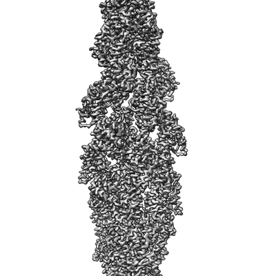 emd_20711.png emd_20711.png | 49 KB | ||
| マスクデータ | emd_20711_msk_1.map | 40.6 MB | マスクマップ | |
| その他 | emd_20711_additional.map.gzemd_20711_additional_1.map.gzemd_20711_half_map_1.map.gzemd_20711_half_map_2.map.gz | 38.2 MB 38.2 MB 7.3 MB 7.3 MB | ||
| アーカイブディレクトリ |  http://ftp.pdbj.org/pub/emdb/structures/EMD-20711ftp://ftp.pdbj.org/pub/emdb/structures/EMD-20711 http://ftp.pdbj.org/pub/emdb/structures/EMD-20711ftp://ftp.pdbj.org/pub/emdb/structures/EMD-20711 | HTTPS FTP |
-関連構造データ


-リンク
| EMDBのページ | EMDB (EBI/PDBe) / EMDataResource |
|---|---|
| 「今月の分子」の関連する項目 |


-マップ
| ファイル | ダウンロード / ファイル: emd_20711.map.gz / 形式: CCP4 / 大きさ: 40.6 MB / タイプ: IMAGE STORED AS FLOATING POINT NUMBER (4 BYTES) | ||||||||||||||||||||||||||||||||||||||||||||||||||||||||||||||||||||
|---|---|---|---|---|---|---|---|---|---|---|---|---|---|---|---|---|---|---|---|---|---|---|---|---|---|---|---|---|---|---|---|---|---|---|---|---|---|---|---|---|---|---|---|---|---|---|---|---|---|---|---|---|---|---|---|---|---|---|---|---|---|---|---|---|---|---|---|---|---|
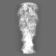
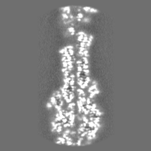
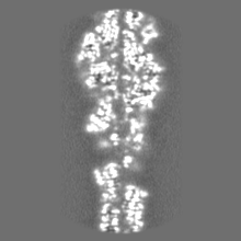
| 注釈 | Helical reconstruction of cofilactin from a partially decorated sample. The map has been masked and sharpened with a b-factor of -215. | ||||||||||||||||||||||||||||||||||||||||||||||||||||||||||||||||||||
| ボクセルのサイズ | X=Y=Z: 1.332 Å | ||||||||||||||||||||||||||||||||||||||||||||||||||||||||||||||||||||
| 密度 |
| ||||||||||||||||||||||||||||||||||||||||||||||||||||||||||||||||||||
| 対称性 | 空間群: 1 | ||||||||||||||||||||||||||||||||||||||||||||||||||||||||||||||||||||
| 詳細 | EMDB XML:
CCP4マップ ヘッダ情報:
| ||||||||||||||||||||||||||||||||||||||||||||||||||||||||||||||||||||
-添付データ
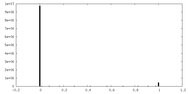
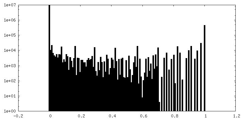
-マスク #1
| ファイル | emd_20711_msk_1.map | ||||||||||||
|---|---|---|---|---|---|---|---|---|---|---|---|---|---|
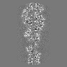
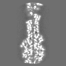
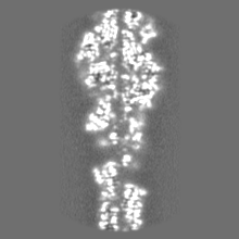
| 投影像・断面図 |
| ||||||||||||
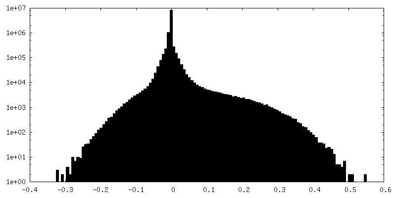
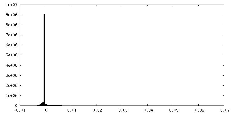
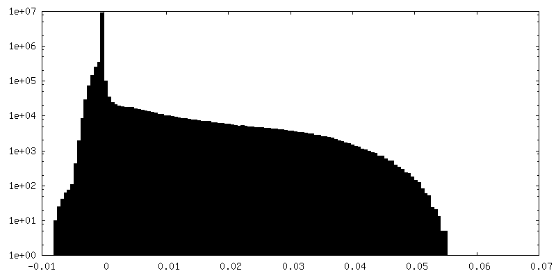
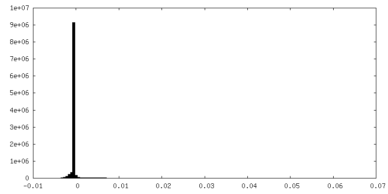
| 密度ヒストグラム |
 Z
Z Y
Y X
X


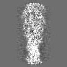
-追加マップ: Unmasked final map.
| ファイル | emd_20711_additional.map | ||||||||||||
|---|---|---|---|---|---|---|---|---|---|---|---|---|---|
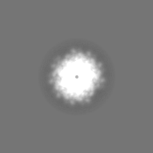
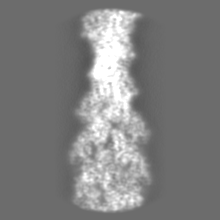
| 注釈 | Unmasked final map. | ||||||||||||
| 投影像・断面図 |
| ||||||||||||
| 密度ヒストグラム |


-追加マップ: Unmasked final map.
| ファイル | emd_20711_additional_1.map | ||||||||||||
|---|---|---|---|---|---|---|---|---|---|---|---|---|---|
| 注釈 | Unmasked final map. | ||||||||||||
| 投影像・断面図 |
| ||||||||||||
| 密度ヒストグラム |


-ハーフマップ: Independently refined half map (2/2) of cofilactin from...
| ファイル | emd_20711_half_map_1.map | ||||||||||||
|---|---|---|---|---|---|---|---|---|---|---|---|---|---|
| 注釈 | Independently refined half map (2/2) of cofilactin from a partially decorated sample. | ||||||||||||
| 投影像・断面図 |
| ||||||||||||
| 密度ヒストグラム |

-ハーフマップ: Independently refined half map (1/2) of cofilactin from...
| ファイル | emd_20711_half_map_2.map | ||||||||||||
|---|---|---|---|---|---|---|---|---|---|---|---|---|---|
| 注釈 | Independently refined half map (1/2) of cofilactin from a partially decorated sample. | ||||||||||||
| 投影像・断面図 |
| ||||||||||||
| 密度ヒストグラム |


- 試料の構成要素
-全体 : Complex of rabbit skeletal actin with human cofilin-1
| 全体 | 名称: Complex of rabbit skeletal actin with human cofilin-1 |
|---|---|
| 要素 |
|
-超分子 #1: Complex of rabbit skeletal actin with human cofilin-1
| 超分子 | 名称: Complex of rabbit skeletal actin with human cofilin-1 タイプ: complex / ID: 1 / 親要素: 0 / 含まれる分子: all |
|---|
-超分子 #2: Skeletal Actin
| 超分子 | 名称: Skeletal Actin / タイプ: complex / ID: 2 / 親要素: 1 / 含まれる分子: #1 |
|---|---|
| 由来(天然) | 生物種: Oryctolagus cuniculus (ウサギ) |
-超分子 #3: Cofilin-1
| 超分子 | 名称: Cofilin-1 / タイプ: complex / ID: 3 / 親要素: 1 / 含まれる分子: #2 |
|---|---|
| 由来(天然) | 生物種: Homo sapiens (ヒト) |
| 組換発現 | 生物種:  Escherichia coli (大腸菌) Escherichia coli (大腸菌) |
-分子 #1: Rabbit Skeletal Actin
| 分子 | 名称: Rabbit Skeletal Actin / タイプ: protein_or_peptide / ID: 1 / 光学異性体: LEVO |
|---|---|
| 由来(天然) | 生物種: Oryctolagus cuniculus (ウサギ) |
| 配列 | 文字列: MCDEDETTAL VCDNGSGLVK AGFAGDDAPR AVFPSIVGRP RHQGVMVGMG QKDSYVGDEA QSKRGILTLK YPIEHGIITN WDDMEKIWHH TFYNELRVAP EEHPTLLTEA PLNPKANREK MTQIMFETFN VPAMYVAIQA VLSLYASGRT TGIVLDSGDG VTHNVPIYEG ...文字列: MCDEDETTAL VCDNGSGLVK AGFAGDDAPR AVFPSIVGRP RHQGVMVGMG QKDSYVGDEA QSKRGILTLK YPIEHGIITN WDDMEKIWHH TFYNELRVAP EEHPTLLTEA PLNPKANREK MTQIMFETFN VPAMYVAIQA VLSLYASGRT TGIVLDSGDG VTHNVPIYEG YALPHAIMRL DLAGRDLTDY LMKILTERGY SFVTTAEREI VRDIKEKLCY VALDFENEMA TAASSSSLEK SYELPDGQVI TIGNERFRCP ETLFQPSFIG MESAGIHETT YNSIMKCDID IRKDLYANNV MSGGTTMYPG IADRMQKEIT ALAPSTMKIK IIAPPERKYS VWIGGSILAS LSTFQQMWIT KQEYDEAGPS IVHRKCF |
-分子 #2: Human Cofilin-1
| 分子 | 名称: Human Cofilin-1 / タイプ: protein_or_peptide / ID: 2 / 光学異性体: LEVO |
|---|---|
| 由来(天然) | 生物種: Homo sapiens (ヒト) |
| 組換発現 | 生物種: Escherichia coli (大腸菌) |
| 配列 | 文字列: MASGVAVSDG VIKVFNDMKV RKSSTPEEVK KRKKAVLFCL SEDKKNIILE EGKEILVGDV GQTVDDPYAT FVKMLPDKDC RYALYDATYE TKESKKEDLV FIFWAPESAP LKSKMIYASS KDAIKKKLTG IKHELQANCY EEVKDRCTLA EKLGGSAVIS LEGKPL |
-実験情報
-構造解析
| 手法 | クライオ電子顕微鏡法 |
|---|---|
 解析 解析 | らせん対称体再構成法 |
| 試料の集合状態 | helical array |
-試料調製
| 緩衝液 | pH: 6.6 |
|---|---|
| グリッド | モデル: Quantifoil R1.2/1.3 / 材質: COPPER |
| 凍結 | 凍結剤: ETHANE / 装置: HOMEMADE PLUNGER |
- 電子顕微鏡法
| 顕微鏡 | FEI TITAN KRIOS |
|---|---|
| 電子線 | 加速電圧: 300 kV / 電子線源: FIELD EMISSION GUN |
| 電子光学系 | 照射モード: SPOT SCAN / 撮影モード: BRIGHT FIELDBright-field microscopy |
| 撮影 | フィルム・検出器のモデル: GATAN K2 SUMMIT (4k x 4k) 平均電子線量: 50.0 e/Å2 |
| 実験機器 |  モデル: Titan Krios / 画像提供: FEI Company |
-画像解析
| Segment selection | 選択した数: 1117338 詳細: Both bare and cofilin-decorated segments were selected and initially refined together |
|---|---|
| 最終 角度割当 | タイプ: NOT APPLICABLE |
| 最終 再構成 | 使用したクラス数: 1 想定した対称性 - らせんパラメータ - Δz: 27.24 Å 想定した対称性 - らせんパラメータ - ΔΦ: -162.5 ° 想定した対称性 - らせんパラメータ - 軸対称性: C1 (非対称) 解像度のタイプ: BY AUTHOR / 解像度: 3.4 Å / 解像度の算出法: FSC 0.143 CUT-OFF / 使用した粒子像数: 558272 |
| FSC曲線 (解像度の算出) | 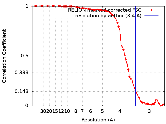 |
