 Movie
Movie Controller

[English] 日本語
 Yorodumi
- PDB-1a0b: HISTIDINE-CONTAINING PHOSPHOTRANSFER DOMAIN OF ARCB FROM ESCHERIC... -
+ Open data

- Basic information
| Entry | Database: PDB / ID: 1a0b | ||||||
|---|---|---|---|---|---|---|---|
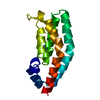
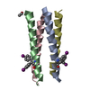
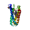
| Title | HISTIDINE-CONTAINING PHOSPHOTRANSFER DOMAIN OF ARCB FROM ESCHERICHIA COLI | ||||||
 Components Components | AEROBIC RESPIRATION CONTROL SENSOR PROTEIN ARCB | ||||||
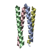
 Keywords Keywords |  HISTIDINE KINASE / PHOSPHOTRANSFER / TWO-COMPONENT SYSTEM / FOUR-HELIX BUNDLE HISTIDINE KINASE / PHOSPHOTRANSFER / TWO-COMPONENT SYSTEM / FOUR-HELIX BUNDLE | ||||||
| Function / homology |  Function and homology information Function and homology informationpeptidyl-histidine phosphorylation / response to oxygen levels / histidine kinase / phosphorelay sensor kinase activity / plasma membrane => GO:0005886 / phosphoprotein phosphatase activity / protein autophosphorylation / regulation of DNA-templated transcription / signal transduction / ATP binding ...peptidyl-histidine phosphorylation / response to oxygen levels / histidine kinase / phosphorelay sensor kinase activity / plasma membrane => GO:0005886 / phosphoprotein phosphatase activity / protein autophosphorylation / regulation of DNA-templated transcription / signal transduction / ATP binding / plasma membrane / cytosolSimilarity search - Function | ||||||
| Biological species |  Escherichia coli (E. coli) Escherichia coli (E. coli) | ||||||
| Method | X-RAY DIFFRACTION / MIR / Resolution: 2.06 Å | ||||||
 Authors Authors | Kato, M. / Mizuno, T. / Shimizu, T. / Hakoshima, T. | ||||||
 Citation Citation | Journal: Cell(Cambridge,Mass.) / Year: 1997 Title: Insights into multistep phosphorelay from the crystal structure of the C-terminal HPt domain of ArcB. Authors: Kato, M. / Mizuno, T. / Shimizu, T. / Hakoshima, T. #1: Journal: Acta Crystallogr.,Sect.D / Year: 1996Title: Crystallization and Preliminary X-Ray Analysis of a Histidine Kinase Domain of the Anaerobic Sensor Protein Arcb from Escherichia Coli Authors: Kato, M. / Ishige, K. / Mizuno, T. / Shimizu, T. / Hakoshima, T. | ||||||
| History |
|
- Structure visualization
| Structure viewer | Molecule: MolmilJmol/JSmol |
|---|
- Downloads & links
-Download
| PDBx/mmCIF format | 1a0b.cif.gz | 38.6 KB | Display | PDBx/mmCIF format |
|---|---|---|---|---|
| PDB format | pdb1a0b.ent.gz | 25.7 KB | Display | PDB format |
| PDBx/mmJSON format | 1a0b.json.gz | Tree view | PDBx/mmJSON format | |
| Others |  Other downloads Other downloads |
-Validation report
| Arichive directory | https://data.pdbj.org/pub/pdb/validation_reports/a0/1a0bftp://data.pdbj.org/pub/pdb/validation_reports/a0/1a0b | HTTPS FTP |
|---|
-Related structure data
| Similar structure data |
|---|


-Links
 PDBj
PDBj


- Assembly
| Deposited unit | 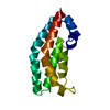
| ||||||||
|---|---|---|---|---|---|---|---|---|---|
| 1 |
| ||||||||
| Unit cell |
|
-Components
| #1: Protein | Mass: 14013.939 Da / Num. of mol.: 1 / Fragment: C-TERMINAL HPT DOMAIN Source method: isolated from a genetically manipulated source Source: (gene. exp.) Escherichia coli (E. coli) / Plasmid: PSU2DH / Cellular location (production host): CYTOPLASM / Production host: Escherichia coli (E. coli) / Strain (production host): K-12 / Variant (production host): DZ225References: UniProt: P22763, UniProt: P0AEC3*PLUS, Transferases; Transferring phosphorus-containing groups; Phosphotransferases with a nitrogenous group as acceptor |
|---|---|
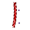
| #2: Chemical | ChemComp-ZN /   Mass: 65.409 Da / Num. of mol.: 1 / Source method: obtained synthetically / Formula: Zn Mass: 65.409 Da / Num. of mol.: 1 / Source method: obtained synthetically / Formula: Zn |
| #3: Water | ChemComp-HOH / Water Mass: 18.015 Da / Num. of mol.: 121 / Source method: isolated from a natural source / Formula: H2O Mass: 18.015 Da / Num. of mol.: 121 / Source method: isolated from a natural source / Formula: H2O |
-Experimental details
-Experiment
| Experiment | Method: X-RAY DIFFRACTION / Number of used crystals: 1 |
|---|
- Sample preparation
| Crystal | Density Matthews: 2.1 Å3/Da / Density % sol: 41 % | ||||||||||||||||||||||||||||||||||||
|---|---|---|---|---|---|---|---|---|---|---|---|---|---|---|---|---|---|---|---|---|---|---|---|---|---|---|---|---|---|---|---|---|---|---|---|---|---|
| Crystal grow | pH: 4.1 Details: PROTEIN WAS CRYSTALLIZED FROM 12.5% PEGMME 550, 5 MM ZNSO4, 50 MM ACETIC ACID/SODIUM ACETATE BUFFER, PH 4.1 | ||||||||||||||||||||||||||||||||||||
| Crystal | *PLUS | ||||||||||||||||||||||||||||||||||||
| Crystal grow | *PLUS Temperature: 277 K / Method: vapor diffusion, hanging dropDetails: Kato, M., (1996) Acta Crystallogr.,Sect.D, 52, 1214. | ||||||||||||||||||||||||||||||||||||
| Components of the solutions | *PLUS
|
-Data collection
| Diffraction | Mean temperature: 277 K |
|---|---|
| Diffraction source | Source: ROTATING ANODE / Type: RIGAKU RUH3R / Wavelength: 1.5418 |
| Detector | Type: RIGAKU RAXIS IIC / Detector: IMAGE PLATE / Date: Jan 1, 1996 / Details: COLLIMATOR |
| Radiation | Monochromator: GRAPHITE(002) / Monochromatic (M) / Laue (L): M / Scattering type: x-ray |
| Radiation wavelength | Wavelength: 1.5418 Å / Relative weight: 1 |
| Reflection | Highest resolution: 2.06 Å / Num. obs: 6999 / % possible obs: 87 % / Observed criterion σ(I): 1 / Redundancy: 3.7 % / Biso Wilson estimate: 20.9 Å2 / Rmerge(I) obs: 0.045 / Net I/σ(I): 17 |
| Reflection shell | Resolution: 2.06→2.25 Å / Redundancy: 3.4 % / Rmerge(I) obs: 0.11 / % possible all: 83 |
| Reflection | *PLUS Num. measured all: 25850 |
- Processing
| Software |
| ||||||||||||||||||||||||||||||||||||||||||||||||||||||||||||||||||||||||||||||||
|---|---|---|---|---|---|---|---|---|---|---|---|---|---|---|---|---|---|---|---|---|---|---|---|---|---|---|---|---|---|---|---|---|---|---|---|---|---|---|---|---|---|---|---|---|---|---|---|---|---|---|---|---|---|---|---|---|---|---|---|---|---|---|---|---|---|---|---|---|---|---|---|---|---|---|---|---|---|---|---|---|---|
| Refinement | Method to determine structure: MIR / Resolution: 2.06→10 Å / Rfactor Rfree error: 0.01 / Data cutoff high absF: 10000000 / Data cutoff low absF: 0.001 / Isotropic thermal model: RESTRAINED / Cross valid method: A POSTERIORI / σ(F): 1
| ||||||||||||||||||||||||||||||||||||||||||||||||||||||||||||||||||||||||||||||||
| Displacement parameters | Biso mean: 28 Å2 | ||||||||||||||||||||||||||||||||||||||||||||||||||||||||||||||||||||||||||||||||
| Refine analyze |
| ||||||||||||||||||||||||||||||||||||||||||||||||||||||||||||||||||||||||||||||||
| Refinement step | Cycle: LAST / Resolution: 2.06→10 Å
| ||||||||||||||||||||||||||||||||||||||||||||||||||||||||||||||||||||||||||||||||
| Refine LS restraints |
| ||||||||||||||||||||||||||||||||||||||||||||||||||||||||||||||||||||||||||||||||
| LS refinement shell | Resolution: 2.06→2.15 Å / Rfactor Rfree error: 0.03 / Total num. of bins used: 8
| ||||||||||||||||||||||||||||||||||||||||||||||||||||||||||||||||||||||||||||||||
| Xplor file |
| ||||||||||||||||||||||||||||||||||||||||||||||||||||||||||||||||||||||||||||||||
| Software | *PLUS Name: X-PLOR / Version: 3.1 / Classification: refinement | ||||||||||||||||||||||||||||||||||||||||||||||||||||||||||||||||||||||||||||||||
| Refine LS restraints | *PLUS
|
