 Movie
Movie Controller

[English] 日本語
 Yorodumi
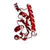
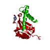
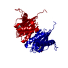
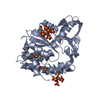
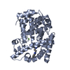
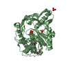
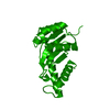
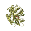
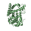
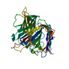
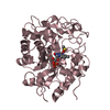
- PDB-1cxq: ATOMIC RESOLUTION ASV INTEGRASE CORE DOMAIN FROM AMMONIUM SULFATE -
+ Open data

- Basic information
| Entry | Database: PDB / ID: 1cxq | ||||||
|---|---|---|---|---|---|---|---|
| Title | ATOMIC RESOLUTION ASV INTEGRASE CORE DOMAIN FROM AMMONIUM SULFATE | ||||||
 Components Components | AVIAN SARCOMA VIRUS INTEGRASE | ||||||
 Keywords Keywords |  TRANSFERASE / MIXED BETA-SHEET SURROUNDED BY ALPHA-HELICES TRANSFERASE / MIXED BETA-SHEET SURROUNDED BY ALPHA-HELICES | ||||||
| Function / homology |  Function and homology informationHydrolases; Acting on peptide bonds (peptidases); Aspartic endopeptidases / ribonuclease H / virion component / DNA integration / RNA-directed DNA polymerase / viral genome integration into host DNA / establishment of integrated proviral latency / RNA-directed DNA polymerase activity / Transferases; Transferring phosphorus-containing groups; Nucleotidyltransferases / RNA-DNA hybrid ribonuclease activity ...Hydrolases; Acting on peptide bonds (peptidases); Aspartic endopeptidases / ribonuclease H / virion component / DNA integration / RNA-directed DNA polymerase / viral genome integration into host DNA / establishment of integrated proviral latency / RNA-directed DNA polymerase activity / Transferases; Transferring phosphorus-containing groups; Nucleotidyltransferases / RNA-DNA hybrid ribonuclease activity / viral nucleocapsid / DNA recombination / Hydrolases; Acting on ester bonds / DNA-directed DNA polymerase / aspartic-type endopeptidase activity / DNA-directed DNA polymerase activity / symbiont entry into host cell / proteolysis / DNA binding / RNA binding / zinc ion binding Function and homology informationHydrolases; Acting on peptide bonds (peptidases); Aspartic endopeptidases / ribonuclease H / virion component / DNA integration / RNA-directed DNA polymerase / viral genome integration into host DNA / establishment of integrated proviral latency / RNA-directed DNA polymerase activity / Transferases; Transferring phosphorus-containing groups; Nucleotidyltransferases / RNA-DNA hybrid ribonuclease activity ...Hydrolases; Acting on peptide bonds (peptidases); Aspartic endopeptidases / ribonuclease H / virion component / DNA integration / RNA-directed DNA polymerase / viral genome integration into host DNA / establishment of integrated proviral latency / RNA-directed DNA polymerase activity / Transferases; Transferring phosphorus-containing groups; Nucleotidyltransferases / RNA-DNA hybrid ribonuclease activity / viral nucleocapsid / DNA recombination / Hydrolases; Acting on ester bonds / DNA-directed DNA polymerase / aspartic-type endopeptidase activity / DNA-directed DNA polymerase activity / symbiont entry into host cell / proteolysis / DNA binding / RNA binding / zinc ion bindingSimilarity search - Function | ||||||
| Biological species |  Avian sarcoma virus Avian sarcoma virus | ||||||
| Method | X-RAY DIFFRACTION / SYNCHROTRON / AB INITIO / Resolution: 1.02 Å | ||||||
 Authors Authors | Lubkowski, J. / Dauter, Z. / Yang, F. / Alexandratos, J. / Merkel, G. / Skalka, A.M. / Wlodawer, A. | ||||||
 Citation Citation | Journal: Biochemistry / Year: 1999 Title: Atomic resolution structures of the core domain of avian sarcoma virus integrase and its D64N mutant. Authors: Lubkowski, J. / Dauter, Z. / Yang, F. / Alexandratos, J. / Merkel, G. / Skalka, A.M. / Wlodawer, A. | ||||||
| History |
|
- Structure visualization
| Structure viewer | Molecule: MolmilJmol/JSmol |
|---|
- Downloads & links
-Download
| PDBx/mmCIF format | 1cxq.cif.gz | 80.7 KB | Display | PDBx/mmCIF format |
|---|---|---|---|---|
| PDB format | pdb1cxq.ent.gz | 60.4 KB | Display | PDB format |
| PDBx/mmJSON format | 1cxq.json.gz | Tree view | PDBx/mmJSON format | |
| Others |  Other downloads Other downloads |
-Validation report
| Arichive directory | https://data.pdbj.org/pub/pdb/validation_reports/cx/1cxqftp://data.pdbj.org/pub/pdb/validation_reports/cx/1cxq | HTTPS FTP |
|---|
-Related structure data


-Links
 PDBj
PDBj
- Assembly
| Deposited unit | 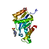
| ||||||||
|---|---|---|---|---|---|---|---|---|---|
| 1 | 
| ||||||||
| Unit cell |
| ||||||||
| Details | The biological assembly is (at least) a dimer constructed from chain A and a symmetry partner generated by the two-fold symmetry operator. |
-Components
| #1: Protein | Mass: 17855.441 Da / Num. of mol.: 1 / Fragment: CATALYTIC CORE DOMAIN / Mutation: INS(P48, L49, R50, E51, N208, L209) Source method: isolated from a genetically manipulated source Source: (gene. exp.) Avian sarcoma virus / Genus: Alpharetrovirus / Plasmid: PRC23IN(52-207) / Production host:  Escherichia coli (E. coli) / References: UniProt: P03354, UniProt: O92956*PLUS Escherichia coli (E. coli) / References: UniProt: P03354, UniProt: O92956*PLUS | ||||
|---|---|---|---|---|---|
| #2: Chemical | ChemComp-EPE / HEPES  Mass: 238.305 Da / Num. of mol.: 1 / Source method: obtained synthetically / Formula: C8H18N2O4S / Comment: pH buffer*YM Mass: 238.305 Da / Num. of mol.: 1 / Source method: obtained synthetically / Formula: C8H18N2O4S / Comment: pH buffer*YM | ||||
| #3: Chemical | Glycerol  Mass: 92.094 Da / Num. of mol.: 2 / Source method: obtained synthetically / Formula: C3H8O3 Mass: 92.094 Da / Num. of mol.: 2 / Source method: obtained synthetically / Formula: C3H8O3#4: Water | ChemComp-HOH / | Water Mass: 18.015 Da / Num. of mol.: 192 / Source method: isolated from a natural source / Formula: H2O Mass: 18.015 Da / Num. of mol.: 192 / Source method: isolated from a natural source / Formula: H2OSequence details | THE APPARENT DISCREPANCY BETWEEN THE SEQUENCE PRESENTED HERE AND THE "POL_RSVP" SEQUENCE IS A ...THE APPARENT DISCREPANC | |
-Experimental details
-Experiment
| Experiment | Method: X-RAY DIFFRACTION / Number of used crystals: 1 |
|---|
- Sample preparation
| Crystal | Density Matthews: 2.41 Å3/Da / Density % sol: 48.93 % | |||||||||||||||||||||||||
|---|---|---|---|---|---|---|---|---|---|---|---|---|---|---|---|---|---|---|---|---|---|---|---|---|---|---|
| Crystal grow | Temperature: 277 K / Method: vapor diffusion, hanging drop / pH: 7.5 Details: 2% Peg 400, 2M Ammonium Sulfate, HEPES pH 7.5, VAPOR DIFFUSION, HANGING DROP, temperature 277.0K | |||||||||||||||||||||||||
| Crystal grow | *PLUS Details: Bujacz, G., (1995) J. Mol. Biol., 253, 333. | |||||||||||||||||||||||||
| Components of the solutions | *PLUS
|
-Data collection
| Diffraction | Mean temperature: 95 K |
|---|---|
| Diffraction source | Source: SYNCHROTRON / Site: NSLS  / Beamline: X9B / Wavelength: 0.98 / Beamline: X9B / Wavelength: 0.98 |
| Detector | Type: MARRESEARCH / Detector: IMAGE PLATE / Date: Mar 21, 1998 |
| Radiation | Protocol: SINGLE WAVELENGTH / Monochromatic (M) / Laue (L): M / Scattering type: x-ray |
| Radiation wavelength | Wavelength: 0.98 Å / Relative weight: 1 |
| Reflection | Resolution: 1.02→20 Å / Num. all: 89063 / Num. obs: 89063 / % possible obs: 100 % / Observed criterion σ(I): -3 / Redundancy: 11.6 % / Biso Wilson estimate: 13.2 Å2 / Rmerge(I) obs: 0.056 / Net I/σ(I): 43.6 |
| Reflection shell | Resolution: 1.02→1.04 Å / Redundancy: 7.7 % / Rmerge(I) obs: 0.029 / Num. unique all: 4376 / % possible all: 99.6 |
| Reflection | *PLUS % possible obs: 100 % / Num. measured all: 1034785 |
| Reflection shell | *PLUS % possible obs: 99.6 % / Mean I/σ(I) obs: 2.9 |
- Processing
| Software |
| |||||||||||||||||||||||||||||||||
|---|---|---|---|---|---|---|---|---|---|---|---|---|---|---|---|---|---|---|---|---|---|---|---|---|---|---|---|---|---|---|---|---|---|---|
| Refinement | Method to determine structure: AB INITIO / Resolution: 1.02→10 Å / Num. parameters: 1238 / Num. restraintsaints: 1519 / σ(F): 0 / Stereochemistry target values: ENGH AND HUBER
| |||||||||||||||||||||||||||||||||
| Refine analyze | Num. disordered residues: 1 / Occupancy sum hydrogen: 953 / Occupancy sum non hydrogen: 1248 | |||||||||||||||||||||||||||||||||
| Refinement step | Cycle: LAST / Resolution: 1.02→10 Å
| |||||||||||||||||||||||||||||||||
| Refine LS restraints |
| |||||||||||||||||||||||||||||||||
| Software | *PLUS Name: SHELXL-96 / Classification: refinement | |||||||||||||||||||||||||||||||||
| Refinement | *PLUS Lowest resolution: 10 Å / σ(F): 0 | |||||||||||||||||||||||||||||||||
| Solvent computation | *PLUS | |||||||||||||||||||||||||||||||||
| Displacement parameters | *PLUS | |||||||||||||||||||||||||||||||||
| Refine LS restraints | *PLUS Type: s_plane_restr / Dev ideal: 0.338 |
