 ムービー
ムービー コントローラー


+ データを開く

- 基本情報
基本情報
| 登録情報 | 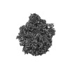 | ||||||||||||
|---|---|---|---|---|---|---|---|---|---|---|---|---|---|
| タイトル | Retapamulin and Capreomycin bound to the 50S subunit | ||||||||||||
 マップデータ マップデータ | |||||||||||||
 試料 試料 |
| ||||||||||||
 キーワード キーワード |  Antibiotic (抗生物質) / RIBOSOME (リボソーム) Antibiotic (抗生物質) / RIBOSOME (リボソーム) | ||||||||||||
| 機能・相同性 |  機能・相同性情報transcriptional attenuation / endoribonuclease inhibitor activity / RNA-binding transcription regulator activity / positive regulation of ribosome biogenesis / negative regulation of cytoplasmic translation / DnaA-L2 complex / translation repressor activity / negative regulation of DNA-templated DNA replication initiation / ribosome assembly / mRNA regulatory element binding translation repressor activity ...transcriptional attenuation / endoribonuclease inhibitor activity / RNA-binding transcription regulator activity / positive regulation of ribosome biogenesis / negative regulation of cytoplasmic translation / DnaA-L2 complex / translation repressor activity / negative regulation of DNA-templated DNA replication initiation / ribosome assembly / mRNA regulatory element binding translation repressor activity / assembly of large subunit precursor of preribosome / cytosolic ribosome assembly / response to reactive oxygen species / regulation of cell growth / DNA-templated transcription termination / response to radiation / ribosomal large subunit assembly / mRNA 5'-UTR binding / large ribosomal subunit / ribosome binding / 5S rRNA binding / large ribosomal subunit rRNA binding / transferase activity / cytosolic large ribosomal subunit / cytoplasmic translation / tRNA binding / negative regulation of translation / rRNA binding / リボソーム / structural constituent of ribosome / 翻訳 (生物学) / response to antibiotic / negative regulation of DNA-templated transcription / mRNA binding / DNA binding / RNA binding / zinc ion binding / 細胞質基質 / 細胞質 機能・相同性情報transcriptional attenuation / endoribonuclease inhibitor activity / RNA-binding transcription regulator activity / positive regulation of ribosome biogenesis / negative regulation of cytoplasmic translation / DnaA-L2 complex / translation repressor activity / negative regulation of DNA-templated DNA replication initiation / ribosome assembly / mRNA regulatory element binding translation repressor activity ...transcriptional attenuation / endoribonuclease inhibitor activity / RNA-binding transcription regulator activity / positive regulation of ribosome biogenesis / negative regulation of cytoplasmic translation / DnaA-L2 complex / translation repressor activity / negative regulation of DNA-templated DNA replication initiation / ribosome assembly / mRNA regulatory element binding translation repressor activity / assembly of large subunit precursor of preribosome / cytosolic ribosome assembly / response to reactive oxygen species / regulation of cell growth / DNA-templated transcription termination / response to radiation / ribosomal large subunit assembly / mRNA 5'-UTR binding / large ribosomal subunit / ribosome binding / 5S rRNA binding / large ribosomal subunit rRNA binding / transferase activity / cytosolic large ribosomal subunit / cytoplasmic translation / tRNA binding / negative regulation of translation / rRNA binding / リボソーム / structural constituent of ribosome / 翻訳 (生物学) / response to antibiotic / negative regulation of DNA-templated transcription / mRNA binding / DNA binding / RNA binding / zinc ion binding / 細胞質基質 / 細胞質類似検索 - 分子機能 | ||||||||||||
| 生物種 |  Escherichia coli BW25113 (大腸菌) / Streptomyces californicus (バクテリア) Escherichia coli BW25113 (大腸菌) / Streptomyces californicus (バクテリア) | ||||||||||||
| 手法 | 単粒子再構成法 / クライオ電子顕微鏡法 / 解像度: 1.83 Å | ||||||||||||
 データ登録者 データ登録者 | Paternoga H / Beckert B / Wilson DN | ||||||||||||
| 資金援助 | European Union, 3件
| ||||||||||||
 引用 引用 | ジャーナル: Nat Struct Mol Biol / 年: 2023 タイトル: Structural conservation of antibiotic interaction with ribosomes. 著者: Helge Paternoga / Caillan Crowe-McAuliffe / Lars V Bock / Timm O Koller / Martino Morici / Bertrand Beckert / Alexander G Myasnikov / Helmut Grubmüller / Jiří Nováček / Daniel N Wilson /    要旨: The ribosome is a major target for clinically used antibiotics, but multidrug resistant pathogenic bacteria are making our current arsenal of antimicrobials obsolete. Here we present cryo-electron- ...The ribosome is a major target for clinically used antibiotics, but multidrug resistant pathogenic bacteria are making our current arsenal of antimicrobials obsolete. Here we present cryo-electron-microscopy structures of 17 distinct compounds from six different antibiotic classes bound to the bacterial ribosome at resolutions ranging from 1.6 to 2.2 Å. The improved resolution enables a precise description of antibiotic-ribosome interactions, encompassing solvent networks that mediate multiple additional interactions between the drugs and their target. Our results reveal a high structural conservation in the binding mode between antibiotics with the same scaffold, including ordered water molecules. Water molecules are visualized within the antibiotic binding sites that are preordered, become ordered in the presence of the drug and that are physically displaced on drug binding. Insight into RNA-ligand interactions will facilitate development of new antimicrobial agents, as well as other RNA-targeting therapies. | ||||||||||||
| 履歴 |
|
- 構造の表示
構造の表示
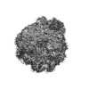
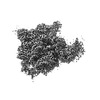
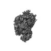
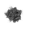
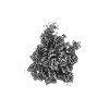
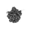
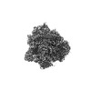
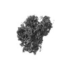
| 添付画像 |
|---|

- ダウンロードとリンク
-EMDBアーカイブ
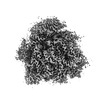
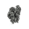
| マップデータ | emd_16613.map.gz | 72.4 MB | EMDBマップデータ形式 | |
|---|---|---|---|---|
| ヘッダ (付随情報) | emd-16613-v30.xmlemd-16613.xml | 55 KB 55 KB | 表示 表示 | EMDBヘッダ |
| 画像 | 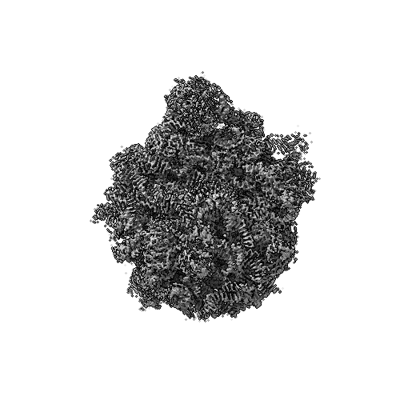 emd_16613.png emd_16613.png | 90 KB | ||
| Filedesc metadata | emd-16613.cif.gz | 11.6 KB | ||
| その他 | emd_16613_additional_1.map.gzemd_16613_half_map_1.map.gzemd_16613_half_map_2.map.gz | 295.2 MB 1.1 GB 1.1 GB | ||
| アーカイブディレクトリ |  http://ftp.pdbj.org/pub/emdb/structures/EMD-16613ftp://ftp.pdbj.org/pub/emdb/structures/EMD-16613 http://ftp.pdbj.org/pub/emdb/structures/EMD-16613ftp://ftp.pdbj.org/pub/emdb/structures/EMD-16613 | HTTPS FTP |
-関連構造データ
| 関連構造データ |  8ceuMC  8ca7C  8caiC 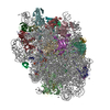 8camC  8cazC  8cepC  8cf1C  8cf8C  8cgdC  8cgiC  8cgjC 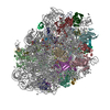 8cgkC 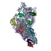 8cgrC 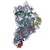 8cguC  8cgvC C: 同じ文献を引用 ( M: このマップから作成された原子モデル |
|---|---|
| 類似構造データ |
-リンク
| EMDBのページ | EMDB (EBI/PDBe) / EMDataResource |
|---|---|
| 「今月の分子」の関連する項目 |


-マップ
| ファイル | ダウンロード / ファイル: emd_16613.map.gz / 形式: CCP4 / 大きさ: 1.4 GB / タイプ: IMAGE STORED AS FLOATING POINT NUMBER (4 BYTES) | ||||||||||||||||||||
|---|---|---|---|---|---|---|---|---|---|---|---|---|---|---|---|---|---|---|---|---|---|
| ボクセルのサイズ | X=Y=Z: 0.72 Å | ||||||||||||||||||||
| 密度 |
| ||||||||||||||||||||
| 対称性 | 空間群: 1 | ||||||||||||||||||||
| 詳細 | EMDB XML:
|
-添付データ
-追加マップ: #1
| ファイル | emd_16613_additional_1.map | ||||||||||||
|---|---|---|---|---|---|---|---|---|---|---|---|---|---|
| 投影像・断面図 |
| ||||||||||||
| 密度ヒストグラム |
 Z
Z Y
Y X
X


-ハーフマップ: #2
| ファイル | emd_16613_half_map_1.map | ||||||||||||
|---|---|---|---|---|---|---|---|---|---|---|---|---|---|
| 投影像・断面図 |
| ||||||||||||
| 密度ヒストグラム |


-ハーフマップ: #1
| ファイル | emd_16613_half_map_2.map | ||||||||||||
|---|---|---|---|---|---|---|---|---|---|---|---|---|---|
| 投影像・断面図 |
| ||||||||||||
| 密度ヒストグラム |


- 試料の構成要素
+全体 : 70S ribosomes with antibiotic cocktail
+超分子 #1: 70S ribosomes with antibiotic cocktail
+分子 #1: Large ribosomal subunit protein bL33
+分子 #2: Large ribosomal subunit protein bL34
+分子 #3: Large ribosomal subunit protein bL35
+分子 #4: Large ribosomal subunit protein bL36A
+分子 #7: Large ribosomal subunit protein uL2
+分子 #8: Large ribosomal subunit protein uL3
+分子 #9: Large ribosomal subunit protein uL4
+分子 #10: Large ribosomal subunit protein uL5
+分子 #11: Large ribosomal subunit protein uL6
+分子 #12: Large ribosomal subunit protein bL9
+分子 #13: Large ribosomal subunit protein uL13
+分子 #14: Large ribosomal subunit protein uL14
+分子 #15: 50S ribosomal protein L15
+分子 #16: Large ribosomal subunit protein uL16
+分子 #17: Large ribosomal subunit protein bL17
+分子 #18: Large ribosomal subunit protein uL18
+分子 #19: Large ribosomal subunit protein bL19
+分子 #20: Large ribosomal subunit protein bL20
+分子 #21: Large ribosomal subunit protein bL21
+分子 #22: Large ribosomal subunit protein uL22
+分子 #23: Large ribosomal subunit protein uL23
+分子 #24: Large ribosomal subunit protein uL24
+分子 #25: 50S ribosomal protein L25
+分子 #26: Large ribosomal subunit protein bL27
+分子 #27: Large ribosomal subunit protein bL28
+分子 #28: Large ribosomal subunit protein uL29
+分子 #29: Large ribosomal subunit protein uL30
+分子 #30: Large ribosomal subunit protein bL32
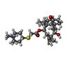
+分子 #31: Capreomycin IA
+分子 #5: 23S rRNA
+分子 #6: 5S rRNA
+分子 #32: ZINC ION
+分子 #33: Retapamulin
+分子 #34: MAGNESIUM ION
+分子 #35: POTASSIUM ION
+分子 #36: water

-実験情報
-構造解析
| 手法 | クライオ電子顕微鏡法 |
|---|---|
 解析 解析 | 単粒子再構成法 |
| 試料の集合状態 | particle |
-試料調製
| 緩衝液 | pH: 7.5 構成要素:
| |||||||||||||||||||||
|---|---|---|---|---|---|---|---|---|---|---|---|---|---|---|---|---|---|---|---|---|---|---|
| グリッド | モデル: Quantifoil R1.2/1.3 / 材質: GOLD / メッシュ: 300 / 支持フィルム - 材質: GRAPHENE | |||||||||||||||||||||
| 凍結 | 凍結剤: ETHANE / チャンバー内湿度: 100 % / チャンバー内温度: 277.15 K |
- 電子顕微鏡法
| 顕微鏡 | FEI TITAN KRIOS |
|---|---|
| 電子線 | 加速電圧: 300 kV / 電子線源: FIELD EMISSION GUN |
| 電子光学系 | 照射モード: FLOOD BEAM / 撮影モード: BRIGHT FIELDBright-field microscopy / 最大 デフォーカス(公称値): 1.0 µm / 最小 デフォーカス(公称値): 0.4 µm |
| 撮影 | フィルム・検出器のモデル: FEI FALCON IV (4k x 4k) 検出モード: COUNTING / 平均露光時間: 4.5 sec. / 平均電子線量: 60.0 e/Å2 |
| 実験機器 |  モデル: Titan Krios / 画像提供: FEI Company |
-画像解析
| 初期モデル | モデルのタイプ: PDB ENTRY PDBモデル - PDB ID: |
|---|---|
| 初期 角度割当 | タイプ: MAXIMUM LIKELIHOOD |
| 最終 角度割当 | タイプ: MAXIMUM LIKELIHOOD |
| 最終 再構成 | 解像度のタイプ: BY AUTHOR / 解像度: 1.83 Å / 解像度の算出法: FSC 0.143 CUT-OFF / 使用した粒子像数: 179724 |

