 Movie
Movie Controller

[English] 日本語
 Yorodumi
- EMDB-1357: Structure of the mitochondrial ATP synthase by electron cryomicro... -
+ Open data

- Basic information
| Entry | Database: EMDB / ID: EMD-1357 | |||||||||
|---|---|---|---|---|---|---|---|---|---|---|
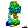
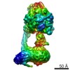
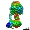
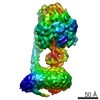
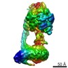
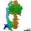
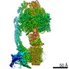
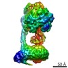
| Title | Structure of the mitochondrial ATP synthase by electron cryomicroscopy. | |||||||||
 Map data Map data | 3-D model of detergent solubilized bovine ATP synthase | |||||||||
 Sample Sample |
| |||||||||
| Biological species |  | |||||||||
| Method | single particle reconstruction / cryo EM / Resolution: 32.0 Å | |||||||||
 Authors Authors | Rubinstein JL / Walker JE / Henderson R | |||||||||
 Citation Citation | Journal: EMBO J / Year: 2003 Title: Structure of the mitochondrial ATP synthase by electron cryomicroscopy. Authors: John L Rubinstein / John E Walker / Richard Henderson /  Abstract: We have determined the structure of intact ATP synthase from bovine heart mitochondria by electron cryomicroscopy of single particles. Docking of an atomic model of the F1-c10 subcomplex into a major ...We have determined the structure of intact ATP synthase from bovine heart mitochondria by electron cryomicroscopy of single particles. Docking of an atomic model of the F1-c10 subcomplex into a major segment of the map has allowed the 32 A resolution density to be interpreted as the F1-ATPase, a central and a peripheral stalk and an FO membrane region that is composed of two domains. One domain of FO corresponds to the ring of c-subunits, and the other probably contains the a-subunit, the transmembrane portion of the b-subunit and the remaining integral membrane proteins of FO. The peripheral stalk wraps around the molecule and connects the apex of F1 to the second domain of FO. The interaction of the peripheral stalk with F1-c10 implies that it binds to a non-catalytic alpha-beta interface in F1 and its inclination where it is not attached to F1 suggests that it has a flexible region that can serve as a stator during both ATP synthesis and ATP hydrolysis. | |||||||||
| History |
|
- Structure visualization
| Movie |
 Movie viewer Movie viewer |
|---|---|
| Structure viewer | EM map: SurfViewMolmilJmol/JSmol |
| Supplemental images |

- Downloads & links
-EMDB archive
| Map data | emd_1357.map.gz | 7.4 MB | EMDB map data format | |
|---|---|---|---|---|
| Header (meta data) | emd-1357-v30.xmlemd-1357.xml | 19.1 KB 19.1 KB | Display Display | EMDB header |
| Images |  1357.gif 1357.gif | 41.3 KB | ||
| Archive directory |  http://ftp.pdbj.org/pub/emdb/structures/EMD-1357ftp://ftp.pdbj.org/pub/emdb/structures/EMD-1357 http://ftp.pdbj.org/pub/emdb/structures/EMD-1357ftp://ftp.pdbj.org/pub/emdb/structures/EMD-1357 | HTTPS FTP |
-Validation report
| Summary document | emd_1357_validation.pdf.gz | 193.2 KB | Display | EMDB validaton report |
|---|---|---|---|---|
| Full document | emd_1357_full_validation.pdf.gz | 192.3 KB | Display | |
| Data in XML | emd_1357_validation.xml.gz | 5.4 KB | Display | |
| Arichive directory | https://ftp.pdbj.org/pub/emdb/validation_reports/EMD-1357ftp://ftp.pdbj.org/pub/emdb/validation_reports/EMD-1357 | HTTPS FTP |
-Related structure data
| Similar structure data |
|---|
-Links
| EMDB pages | EMDB (EBI/PDBe) / EMDataResource |
|---|
-Map
| File | Download / File: emd_1357.map.gz / Format: CCP4 / Size: 7.8 MB / Type: IMAGE STORED AS FLOATING POINT NUMBER (4 BYTES) | ||||||||||||||||||||||||||||||||||||||||||||||||||||||||||||||||||||
|---|---|---|---|---|---|---|---|---|---|---|---|---|---|---|---|---|---|---|---|---|---|---|---|---|---|---|---|---|---|---|---|---|---|---|---|---|---|---|---|---|---|---|---|---|---|---|---|---|---|---|---|---|---|---|---|---|---|---|---|---|---|---|---|---|---|---|---|---|---|
| Annotation | 3-D model of detergent solubilized bovine ATP synthase | ||||||||||||||||||||||||||||||||||||||||||||||||||||||||||||||||||||
| Voxel size | X=Y=Z: 2.8 Å | ||||||||||||||||||||||||||||||||||||||||||||||||||||||||||||||||||||
| Density |
| ||||||||||||||||||||||||||||||||||||||||||||||||||||||||||||||||||||
| Symmetry | Space group: 1 | ||||||||||||||||||||||||||||||||||||||||||||||||||||||||||||||||||||
| Details | EMDB XML:
CCP4 map header:
| ||||||||||||||||||||||||||||||||||||||||||||||||||||||||||||||||||||
-Supplemental data
- Sample components
+Entire : Bovine mitochondrial ATP synthase
+Supramolecule #1000: Bovine mitochondrial ATP synthase
+Macromolecule #1: alpha
+Macromolecule #2: beta
+Macromolecule #3: gamma
+Macromolecule #4: delta
+Macromolecule #5: epsilon
+Macromolecule #6: a
+Macromolecule #7: b
+Macromolecule #8: c
+Macromolecule #9: d
+Macromolecule #10: e
+Macromolecule #11: f
+Macromolecule #12: g
+Macromolecule #13: F6
+Macromolecule #14: OSCP
+Macromolecule #15: A6L
-Experimental details
-Structure determination
| Method | cryo EM |
|---|---|
 Processing Processing | single particle reconstruction |
| Aggregation state | particle |
-Sample preparation
| Concentration | 3 mg/mL |
|---|---|
| Buffer | pH: 8 Details: Tris-HCl, 20 mM Sucrose, 50 mM Magnesium Sulfate, 2 mM EDTA, 1 mM ADP, 2 mM Brij-35, 0.05%(w/v) |
| Grid | Details: 400 mesh grid coated with a perforated carbon film |
| Vitrification | Cryogen name: ETHANE / Chamber humidity: 100 % / Instrument: HOMEMADE PLUNGER Details: Vitrification instrument: Talmon plunger. Carried out in coldroom Method: Sample applied from one side and blotted from same side |
- Electron microscopy
| Microscope | FEI TECNAI F20 |
|---|---|
| Alignment procedure | Legacy - Astigmatism: corrected at 175kx magnification |
| Details | Digitized with Zeiss SCAI scanner |
| Date | Jan 1, 2003 |
| Image recording | Category: FILM / Film or detector model: KODAK SO-163 FILM / Digitization - Scanner: ZEISS SCAI / Digitization - Sampling interval: 7 µm / Number real images: 101 / Average electron dose: 10 e/Å2 / Bits/pixel: 8 |
| Electron beam | Acceleration voltage: 200 kV / Electron source:  FIELD EMISSION GUN FIELD EMISSION GUN |
| Electron optics | Illumination mode: FLOOD BEAM / Imaging mode: BRIGHT FIELD / Cs: 2.0 mm / Nominal defocus max: 6.0 µm / Nominal defocus min: 3.0 µm / Nominal magnification: 50000 |
| Sample stage | Specimen holder: Side entry liquid nitrogen / Specimen holder model: GATAN LIQUID NITROGEN |
| Experimental equipment |  Model: Tecnai F20 / Image courtesy: FEI Company |
-Image processing
| Details | Particles manually selected. |
|---|---|
| CTF correction | Details: See publication |
| Final reconstruction | Applied symmetry - Point group: C1 (asymmetric) / Algorithm: OTHER / Resolution.type: BY AUTHOR / Resolution: 32.0 Å / Resolution method: FSC 0.143 CUT-OFF / Software - Name: IMAGIC, FREALIGN, ROTAN / Number images used: 5984 |
-Atomic model buiding 1
| Software | Name: O |
|---|---|
| Details | Protocol: Eye. Multiple pdb files fit. See publication. |
| Refinement | Protocol: RIGID BODY FIT |
