- EMDB-12179: Staphylococcus aureus 30S ribosomal subunit in presence of spermi... -
+
データを開く
IDまたはキーワード:
読み込み中...
-
基本情報
登録情報
データベース: EMDB / ID: EMD-12179
タイトル
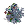
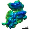
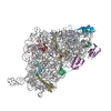
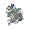
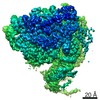
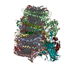
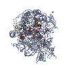
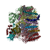
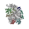
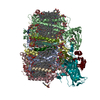
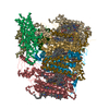
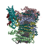
Staphylococcus aureus 30S ribosomal subunit in presence of spermidine (head only)
マップデータ
試料
複合体: Staphylococcus aureus 30S ribosomal subunit in presence of spermidine (head only)
RNA: 16S ribosomal RNA16SリボソームRNA
タンパク質・ペプチド: 30S ribosomal protein S2
タンパク質・ペプチド: 30S ribosomal protein S3
タンパク質・ペプチド: 30S ribosomal protein S7
タンパク質・ペプチド: 30S ribosomal protein S9
タンパク質・ペプチド: 30S ribosomal protein S10
タンパク質・ペプチド: 30S ribosomal protein S13
タンパク質・ペプチド: 30S ribosomal protein S14 type Zリボソーム
タンパク質・ペプチド: 30S ribosomal protein S19
機能・相同性
機能・相同性情報
ribosomal small subunit assembly / small ribosomal subunit / cytosolic small ribosomal subunit / tRNA binding / rRNA binding / リボソーム / structural constituent of ribosome / ribonucleoprotein complex / 翻訳 (生物学) / mRNA binding ...ribosomal small subunit assembly / small ribosomal subunit / cytosolic small ribosomal subunit / tRNA binding / rRNA binding / リボソーム / structural constituent of ribosome / ribonucleoprotein complex / 翻訳 (生物学) / mRNA binding / RNA binding / zinc ion binding / 細胞質基質 類似検索 - 分子機能
Ribosomal protein S14, type Z / Ribosomal protein S14/S29 / Ribosomal protein S3, bacterial-type / Ribosomal protein S19, bacterial-type / Ribosomal protein S7, bacterial/organellar-type / Ribosomal protein S13, bacterial-type / Ribosomal protein S9, bacterial/plastid / Ribosomal protein S2, bacteria/mitochondria/plastid / Ribosomal protein S2 signature 2. / KHドメイン ...Ribosomal protein S14, type Z / Ribosomal protein S14/S29 / Ribosomal protein S3, bacterial-type / Ribosomal protein S19, bacterial-type / Ribosomal protein S7, bacterial/organellar-type / Ribosomal protein S13, bacterial-type / Ribosomal protein S9, bacterial/plastid / Ribosomal protein S2, bacteria/mitochondria/plastid / Ribosomal protein S2 signature 2. / KHドメイン / K homology RNA-binding domain / Ribosomal protein S3, conserved site / Ribosomal protein S3 signature. / Ribosomal protein S10, conserved site / Ribosomal protein S10 signature. / Ribosomal protein S14, conserved site / Ribosomal protein S14 signature. / Ribosomal protein S2 signature 1. / Ribosomal protein S2, conserved site / Ribosomal protein S2 / Ribosomal protein S2, flavodoxin-like domain superfamily / KHドメイン / Ribosomal protein S2 / Type-2 KH domain profile. / K Homology domain, type 2 / Ribosomal protein S3, C-terminal / Ribosomal protein S3, C-terminal domain / Ribosomal protein S3, C-terminal domain superfamily / Ribosomal protein S15/S19, conserved site / Ribosomal protein S19 signature. / Ribosomal protein S10 / Ribosomal protein S19/S15 / Ribosomal protein S19/S15, superfamily / Ribosomal protein S19 / Ribosomal protein S7, conserved site / Ribosomal protein S7 signature. / K homology domain superfamily, prokaryotic type / Ribosomal protein S13, conserved site / Ribosomal protein S13 signature. / Ribosomal protein S13 / 30s ribosomal protein S13, C-terminal / Ribosomal protein S13/S18 / Ribosomal protein S13 family profile. / Ribosomal protein S14 / Ribosomal protein S14p/S29e / K homology domain-like, alpha/beta / Ribosomal protein S10p/S20e / Ribosomal protein S10 domain / Ribosomal protein S10 domain superfamily / Ribosomal protein S10p/S20e / Ribosomal protein S9, conserved site / Ribosomal protein S9 signature. / Ribosomal protein S13-like, H2TH / Ribosomal protein S5/S7 / Ribosomal protein S7 domain / Ribosomal protein S7 domain superfamily / Ribosomal protein S7p/S5e / Ribosomal protein S9 / Ribosomal protein S9/S16 / Ribosomal protein S5 domain 2-type fold, subgroup / Ribosomal protein S5 domain 2-type fold 類似検索 - ドメイン・相同性
Small ribosomal subunit protein uS7 / Small ribosomal subunit protein uS19 / Small ribosomal subunit protein uS3 / Small ribosomal subunit protein uS14B / Small ribosomal subunit protein uS13 / Small ribosomal subunit protein uS9 / Small ribosomal subunit protein uS2 / Small ribosomal subunit protein uS10 類似検索 - 構成要素
ジャーナル: Front Mol Biosci / 年: 2021 タイトル: Stabilization of Ribosomal RNA of the Small Subunit by Spermidine in . 著者: Margarita Belinite / Iskander Khusainov / Heddy Soufari / Stefano Marzi / Pascale Romby / Marat Yusupov / Yaser Hashem / 要旨: Cryo-electron microscopy is now used as a method of choice in structural biology for studying protein synthesis, a process mediated by the ribosome machinery. In order to achieve high-resolution ...Cryo-electron microscopy is now used as a method of choice in structural biology for studying protein synthesis, a process mediated by the ribosome machinery. In order to achieve high-resolution structures using this approach, one needs to obtain homogeneous and stable samples, which requires optimization of ribosome purification in a species-dependent manner. This is especially critical for the bacterial small ribosomal subunit that tends to be unstable in the absence of ligands. Here, we report a protocol for purification of stable 30 S from the Gram-positive bacterium and its cryo-EM structures: in presence of spermidine at a resolution ranging between 3.4 and 3.6 Å and in its absence at 5.3 Å. Using biochemical characterization and cryo-EM, we demonstrate the importance of spermidine for stabilization of the 30 S preserving favorable conformation of the helix 44.
 ムービー
ムービー コントローラー


 データを開く
 基本情報
基本情報 マップデータ
 試料
試料 機能・相同性情報
 ribosomal small subunit assembly / small ribosomal subunit / cytosolic small ribosomal subunit /
 データ登録者
 フランス, 2件
  引用
引用
 構造の表示
構造の表示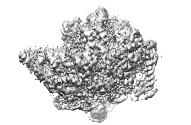
 ダウンロードとリンク
 emd_12179.png
 http://ftp.pdbj.org/pub/emdb/structures/EMD-12179
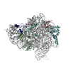


 試料の構成要素
 解析
解析 電子顕微鏡法