 ムービー
ムービー コントローラー


+ データを開く

- 基本情報
基本情報
| 登録情報 | データベース: EMDB / ID: EMD-11697 | |||||||||
|---|---|---|---|---|---|---|---|---|---|---|
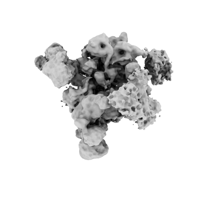
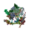
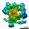
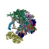
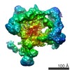
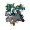
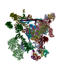
| タイトル | Human pre-Bact-2 spliceosome | |||||||||
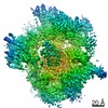
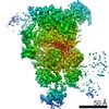
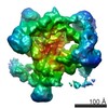
 マップデータ マップデータ | Unmasked/unsharpened map of the human pre-Bact-2 spliceosome. | |||||||||
 試料 試料 |
| |||||||||
| 機能・相同性 |  機能・相同性情報 機能・相同性情報RES complex /  microfibril / somatic diversification of immunoglobulins / post-mRNA release spliceosomal complex / U11/U12 snRNP / regulation of retinoic acid receptor signaling pathway / 3'-5' RNA helicase activity / snRNP binding / U2 snRNP binding / U7 snRNA binding ...RES complex / microfibril / somatic diversification of immunoglobulins / post-mRNA release spliceosomal complex / U11/U12 snRNP / regulation of retinoic acid receptor signaling pathway / 3'-5' RNA helicase activity / snRNP binding / U2 snRNP binding / U7 snRNA binding / histone pre-mRNA DCP binding / generation of catalytic spliceosome for first transesterification step / U7 snRNP / regulation of vitamin D receptor signaling pathway / B-WICH complex / histone pre-mRNA 3'end processing complex / cis assembly of pre-catalytic spliceosome / SLBP independent Processing of Histone Pre-mRNAs / SLBP Dependent Processing of Replication-Dependent Histone Pre-mRNAs / embryonic brain development / splicing factor binding / spliceosome conformational change to release U4 (or U4atac) and U1 (or U11) / protein methylation / miRNA processing / U12-type spliceosomal complex / methylosome / nuclear retinoic acid receptor binding / Prp19 complex / 7-methylguanosine cap hypermethylation / positive regulation of androgen receptor activity / poly(A) binding / U1 snRNP binding / pICln-Sm protein complex / mRNA 3'-end processing / : / blastocyst formation / pre-mRNA binding / U2-type catalytic step 1 spliceosome / RNA splicing, via transesterification reactions / small nuclear ribonucleoprotein complex / sno(s)RNA-containing ribonucleoprotein complex / P granule / SMN-Sm protein complex / regulation of mRNA splicing, via spliceosome / spliceosomal tri-snRNP complex / telomerase holoenzyme complex / mRNA cis splicing, via spliceosome / U2-type spliceosomal complex / positive regulation by host of viral transcription / telomerase RNA binding / U2-type precatalytic spliceosome / commitment complex / positive regulation of vitamin D receptor signaling pathway / Transport of Mature mRNA derived from an Intron-Containing Transcript / transcription regulator inhibitor activity / U2-type prespliceosome assembly / U2-type catalytic step 2 spliceosome / nuclear vitamin D receptor binding / U4 snRNP / Notch binding / Regulation of gene expression in late stage (branching morphogenesis) pancreatic bud precursor cells / RUNX3 regulates NOTCH signaling / SAGA complex / positive regulation of mRNA splicing, via spliceosome / U2 snRNP / Basigin interactions / RNA Polymerase II Transcription Termination / NOTCH4 Intracellular Domain Regulates Transcription / RHOBTB1 GTPase cycle / positive regulation of transcription by RNA polymerase III / U1 snRNP / ubiquitin-ubiquitin ligase activity / positive regulation of protein targeting to mitochondrion / WD40-repeat domain binding / NOTCH3 Intracellular Domain Regulates Transcription / positive regulation of neurogenesis / U2-type prespliceosome / K63-linked polyubiquitin modification-dependent protein binding / nuclear androgen receptor binding / cyclosporin A binding / precatalytic spliceosome / positive regulation of transcription by RNA polymerase I / spliceosomal complex assembly / Notch-HLH transcription pathway / Formation of paraxial mesoderm / regulation of alternative mRNA splicing, via spliceosome / mRNA Splicing - Minor Pathway / positive regulation of transforming growth factor beta receptor signaling pathway / SMAD binding / blastocyst development / regulation of RNA splicing / mRNA 3'-splice site recognition / protein localization to nucleus / spliceosomal tri-snRNP complex assembly / intrinsic apoptotic signaling pathway in response to DNA damage by p53 class mediator / retinoic acid receptor signaling pathway / U5 snRNA binding / positive regulation of G1/S transition of mitotic cell cycle / U5 snRNP / transcription-coupled nucleotide-excision repair microfibril / somatic diversification of immunoglobulins / post-mRNA release spliceosomal complex / U11/U12 snRNP / regulation of retinoic acid receptor signaling pathway / 3'-5' RNA helicase activity / snRNP binding / U2 snRNP binding / U7 snRNA binding ...RES complex / microfibril / somatic diversification of immunoglobulins / post-mRNA release spliceosomal complex / U11/U12 snRNP / regulation of retinoic acid receptor signaling pathway / 3'-5' RNA helicase activity / snRNP binding / U2 snRNP binding / U7 snRNA binding / histone pre-mRNA DCP binding / generation of catalytic spliceosome for first transesterification step / U7 snRNP / regulation of vitamin D receptor signaling pathway / B-WICH complex / histone pre-mRNA 3'end processing complex / cis assembly of pre-catalytic spliceosome / SLBP independent Processing of Histone Pre-mRNAs / SLBP Dependent Processing of Replication-Dependent Histone Pre-mRNAs / embryonic brain development / splicing factor binding / spliceosome conformational change to release U4 (or U4atac) and U1 (or U11) / protein methylation / miRNA processing / U12-type spliceosomal complex / methylosome / nuclear retinoic acid receptor binding / Prp19 complex / 7-methylguanosine cap hypermethylation / positive regulation of androgen receptor activity / poly(A) binding / U1 snRNP binding / pICln-Sm protein complex / mRNA 3'-end processing / : / blastocyst formation / pre-mRNA binding / U2-type catalytic step 1 spliceosome / RNA splicing, via transesterification reactions / small nuclear ribonucleoprotein complex / sno(s)RNA-containing ribonucleoprotein complex / P granule / SMN-Sm protein complex / regulation of mRNA splicing, via spliceosome / spliceosomal tri-snRNP complex / telomerase holoenzyme complex / mRNA cis splicing, via spliceosome / U2-type spliceosomal complex / positive regulation by host of viral transcription / telomerase RNA binding / U2-type precatalytic spliceosome / commitment complex / positive regulation of vitamin D receptor signaling pathway / Transport of Mature mRNA derived from an Intron-Containing Transcript / transcription regulator inhibitor activity / U2-type prespliceosome assembly / U2-type catalytic step 2 spliceosome / nuclear vitamin D receptor binding / U4 snRNP / Notch binding / Regulation of gene expression in late stage (branching morphogenesis) pancreatic bud precursor cells / RUNX3 regulates NOTCH signaling / SAGA complex / positive regulation of mRNA splicing, via spliceosome / U2 snRNP / Basigin interactions / RNA Polymerase II Transcription Termination / NOTCH4 Intracellular Domain Regulates Transcription / RHOBTB1 GTPase cycle / positive regulation of transcription by RNA polymerase III / U1 snRNP / ubiquitin-ubiquitin ligase activity / positive regulation of protein targeting to mitochondrion / WD40-repeat domain binding / NOTCH3 Intracellular Domain Regulates Transcription / positive regulation of neurogenesis / U2-type prespliceosome / K63-linked polyubiquitin modification-dependent protein binding / nuclear androgen receptor binding / cyclosporin A binding / precatalytic spliceosome / positive regulation of transcription by RNA polymerase I / spliceosomal complex assembly / Notch-HLH transcription pathway / Formation of paraxial mesoderm / regulation of alternative mRNA splicing, via spliceosome / mRNA Splicing - Minor Pathway / positive regulation of transforming growth factor beta receptor signaling pathway / SMAD binding / blastocyst development / regulation of RNA splicing / mRNA 3'-splice site recognition / protein localization to nucleus / spliceosomal tri-snRNP complex assembly / intrinsic apoptotic signaling pathway in response to DNA damage by p53 class mediator / retinoic acid receptor signaling pathway / U5 snRNA binding / positive regulation of G1/S transition of mitotic cell cycle / U5 snRNP / transcription-coupled nucleotide-excision repair類似検索 - 分子機能 | |||||||||
| 生物種 |  Homo sapiens (ヒト) / synthetic construct (人工物) / Human (ヒト) Homo sapiens (ヒト) / synthetic construct (人工物) / Human (ヒト) | |||||||||
| 手法 | 単粒子再構成法 / クライオ電子顕微鏡法 / 解像度: 8.0 Å | |||||||||
 データ登録者 データ登録者 | Townsend C / Kastner B / Leelaram MN / Bertram K / Stark H / Luehrmann R | |||||||||
| 資金援助 |  ドイツ, 1件 ドイツ, 1件
| |||||||||
 引用 引用 | ジャーナル: Science / 年: 2020 タイトル: Mechanism of protein-guided folding of the active site U2/U6 RNA during spliceosome activation. 著者: Cole Townsend / Majety N Leelaram / Dmitry E Agafonov / Olexandr Dybkov / Cindy L Will / Karl Bertram / Henning Urlaub / Berthold Kastner / Holger Stark / Reinhard Lührmann / 要旨: Spliceosome activation involves extensive protein and RNA rearrangements that lead to formation of a catalytically active U2/U6 RNA structure. At present, little is known about the assembly pathway ...Spliceosome activation involves extensive protein and RNA rearrangements that lead to formation of a catalytically active U2/U6 RNA structure. At present, little is known about the assembly pathway of the latter and the mechanism whereby proteins aid its proper folding. Here, we report the cryo-electron microscopy structures of two human, activated spliceosome precursors (that is, pre-B complexes) at core resolutions of 3.9 and 4.2 angstroms. These structures elucidate the order of the numerous protein exchanges that occur during activation, the mutually exclusive interactions that ensure the correct order of ribonucleoprotein rearrangements needed to form the U2/U6 catalytic RNA, and the stepwise folding pathway of the latter. Structural comparisons with mature B complexes reveal the molecular mechanism whereby a conformational change in the scaffold protein PRP8 facilitates final three-dimensional folding of the U2/U6 catalytic RNA. | |||||||||
| 履歴 |
|
- 構造の表示
構造の表示
| ムービー |
ムービービューア |
|---|---|
| 構造ビューア | EMマップ: SurfViewMolmilJmol/JSmol |
| 添付画像 |
- ダウンロードとリンク
-EMDBアーカイブ
| マップデータ | emd_11697.map.gz | 171.3 MB | EMDBマップデータ形式 | |
|---|---|---|---|---|
| ヘッダ (付随情報) | emd-11697-v30.xmlemd-11697.xml | 80.2 KB 80.2 KB | 表示 表示 | EMDBヘッダ |
| 画像 |  emd_11697.png emd_11697.png | 39.9 KB | ||
| アーカイブディレクトリ |  http://ftp.pdbj.org/pub/emdb/structures/EMD-11697ftp://ftp.pdbj.org/pub/emdb/structures/EMD-11697 http://ftp.pdbj.org/pub/emdb/structures/EMD-11697ftp://ftp.pdbj.org/pub/emdb/structures/EMD-11697 | HTTPS FTP |
-関連構造データ
| 関連構造データ | 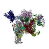 7abiMC 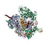 7aavC 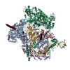 7abfC 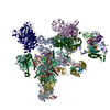 7abgC 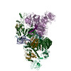 7abhC C: 同じ文献を引用 ( M: このマップから作成された原子モデル |
|---|---|
| 類似構造データ | |
| 電子顕微鏡画像生データ | EMPIAR-10616 (タイトル: Cryo-EM dataset of human pre-Bact spliceosome / Data size: 584.5 Data #1: Motion-corrected micrographs (without dose-weighting) of human pre-Bact spliceosome [micrographs - single frame] Data #2: Motion-corrected micrographs (with dose-weighting) of human pre-Bact spliceosome [micrographs - single frame]) |


-リンク
| EMDBのページ | EMDB (EBI/PDBe) / EMDataResource |
|---|---|
| 「今月の分子」の関連する項目 |


-マップ
| ファイル | ダウンロード / ファイル: emd_11697.map.gz / 形式: CCP4 / 大きさ: 216 MB / タイプ: IMAGE STORED AS FLOATING POINT NUMBER (4 BYTES) | ||||||||||||||||||||||||||||||||||||||||||||||||||||||||||||
|---|---|---|---|---|---|---|---|---|---|---|---|---|---|---|---|---|---|---|---|---|---|---|---|---|---|---|---|---|---|---|---|---|---|---|---|---|---|---|---|---|---|---|---|---|---|---|---|---|---|---|---|---|---|---|---|---|---|---|---|---|---|
| 注釈 | Unmasked/unsharpened map of the human pre-Bact-2 spliceosome. | ||||||||||||||||||||||||||||||||||||||||||||||||||||||||||||
| ボクセルのサイズ | X=Y=Z: 1.16 Å | ||||||||||||||||||||||||||||||||||||||||||||||||||||||||||||
| 密度 |
| ||||||||||||||||||||||||||||||||||||||||||||||||||||||||||||
| 対称性 | 空間群: 1 | ||||||||||||||||||||||||||||||||||||||||||||||||||||||||||||
| 詳細 | EMDB XML:
CCP4マップ ヘッダ情報:
| ||||||||||||||||||||||||||||||||||||||||||||||||||||||||||||
-添付データ
- 試料の構成要素
+全体 : Human pre-Bact-2 spliceosome
+超分子 #1: Human pre-Bact-2 spliceosome
+超分子 #2: Human pre-Bact-2 spliceosome
+超分子 #3: MINX M3 pre-mRNA
+分子 #1: U5 small nuclear ribonucleoprotein 40 kDa protein
+分子 #2: Splicing factor 3B subunit 3
+分子 #3: Small nuclear ribonucleoprotein F
+分子 #4: U2 small nuclear ribonucleoprotein A'
+分子 #5: Splicing factor 3B subunit 4
+分子 #6: Small nuclear ribonucleoprotein G
+分子 #7: Intron-binding protein aquarius
+分子 #8: Splicing factor 3B subunit 5
+分子 #10: U2 small nuclear ribonucleoprotein B''
+分子 #11: Splicing factor 3B subunit 6
+分子 #12: Ubiquitin-like protein 5
+分子 #13: U5 small nuclear ribonucleoprotein 200 kDa helicase
+分子 #14: SNW domain-containing protein 1
+分子 #15: BUD13 homolog
+分子 #16: Smad nuclear-interacting protein 1
+分子 #17: Protein BUD31 homolog
+分子 #18: RNA-binding motif protein, X-linked 2
+分子 #19: Cell division cycle 5-like protein
+分子 #20: Zinc finger matrin-type protein 2
+分子 #21: Beta-catenin-like protein 1
+分子 #22: 116 kDa U5 small nuclear ribonucleoprotein component
+分子 #23: Spliceosome-associated protein CWC15 homolog
+分子 #24: Serine/arginine repetitive matrix protein 1
+分子 #25: DNA/RNA-binding protein KIN17
+分子 #26: Pre-mRNA-splicing factor SYF1
+分子 #27: Microfibrillar-associated protein 1
+分子 #28: Crooked neck-like protein 1
+分子 #29: PHD finger-like domain-containing protein 5A
+分子 #31: Pleiotropic regulator 1
+分子 #32: Small nuclear ribonucleoprotein Sm D2
+分子 #33: Peptidyl-prolyl cis-trans isomerase E
+分子 #34: RING-type E3 ubiquitin-protein ligase PPIL2
+分子 #35: Small nuclear ribonucleoprotein-associated proteins B and B'
+分子 #37: Small nuclear ribonucleoprotein Sm D3
+分子 #38: Pre-mRNA-processing-splicing factor 8
+分子 #39: Pre-mRNA-processing factor 17
+分子 #40: Small nuclear ribonucleoprotein E
+分子 #41: Pre-mRNA-splicing factor 38A
+分子 #42: Small nuclear ribonucleoprotein Sm D1
+分子 #43: Pre-mRNA-splicing factor RBM22
+分子 #45: Splicing factor 3A subunit 1
+分子 #46: Splicing factor 3A subunit 2
+分子 #47: Splicing factor 3A subunit 3
+分子 #48: Splicing factor 3B subunit 1
+分子 #49: Splicing factor 3B subunit 2
+分子 #9: U6 snRNA
+分子 #30: U2 snRNA
+分子 #36: MINX M3 pre-mRNA
+分子 #44: U5 snRNA
+分子 #50: GUANOSINE-5'-TRIPHOSPHATE

+分子 #51: MAGNESIUM ION
+分子 #52: INOSITOL HEXAKISPHOSPHATE

-実験情報
-構造解析
| 手法 | クライオ電子顕微鏡法 |
|---|---|
 解析 解析 | 単粒子再構成法 |
| 試料の集合状態 | particle |
-試料調製
| 緩衝液 | pH: 7.9 |
|---|---|
| グリッド | モデル: Quantifoil R3.5/1 / 材質: COPPER |
| 凍結 | 凍結剤: ETHANE |
- 電子顕微鏡法
| 顕微鏡 | FEI TITAN KRIOS |
|---|---|
| 電子線 | 加速電圧: 300 kV / 電子線源: FIELD EMISSION GUN |
| 電子光学系 | 照射モード: SPOT SCAN / 撮影モード: BRIGHT FIELDBright-field microscopy |
| 撮影 | フィルム・検出器のモデル: FEI FALCON III (4k x 4k) 検出モード: INTEGRATING / 平均露光時間: 1.0 sec. / 平均電子線量: 2.27 e/Å2 |
| 実験機器 |  モデル: Titan Krios / 画像提供: FEI Company |
-画像解析
| 初期モデル | モデルのタイプ: OTHER / 詳細: cryoSPARC ab initio |
|---|---|
| 初期 角度割当 | タイプ: MAXIMUM LIKELIHOOD |
| 最終 角度割当 | タイプ: MAXIMUM LIKELIHOOD |
| 最終 再構成 | 解像度のタイプ: BY AUTHOR / 解像度: 8.0 Å / 解像度の算出法: FSC 0.143 CUT-OFF / ソフトウェア - 名称: RELION (ver. 3.0) / 使用した粒子像数: 39336 |
-原子モデル構築 1
| 精密化 | 空間: REAL / プロトコル: RIGID BODY FIT |
|---|---|
| 得られたモデル | PDB-7abi: |
