 Movie
Movie Controller


+ Open data

- Basic information
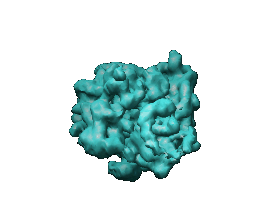
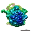
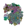
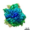
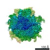
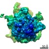
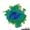
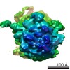
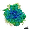
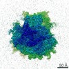
| Entry | Database: EMDB / ID: EMD-1122 | |||||||||
|---|---|---|---|---|---|---|---|---|---|---|
| Title | Visualizing tmRNA entry into a stalled ribosome. | |||||||||
 Map data Map data | this is a ribosome consisting 70S + Psite tRNA + mRNA + EF-Tu.GDP + alanylated tmRNA + SmpB + Kirromycin + S1 | |||||||||
 Sample Sample |
| |||||||||
| Function / homology |  Function and homology information Function and homology informationtrans-translation / translation elongation factor activity / GTPase activity / GTP binding / RNA binding / cytosol Similarity search - Function | |||||||||
| Biological species |   Thermus thermophilus HB8 (bacteria) Thermus thermophilus HB8 (bacteria) | |||||||||
| Method | single particle reconstruction / cryo EM / Resolution: 13.0 Å | |||||||||
 Authors Authors | Valle M / Gillet R / Kaur S / Henne A / Ramakrishnan V / Frank J | |||||||||
 Citation Citation | Journal: Science / Year: 2003 Title: Visualizing tmRNA entry into a stalled ribosome. Authors: Mikel Valle / Reynald Gillet / Sukhjit Kaur / Anke Henne / V Ramakrishnan / Joachim Frank /  Abstract: Bacterial ribosomes stalled on defective messenger RNAs (mRNAs) are rescued by tmRNA, an approximately 300-nucleotide-long molecule that functions as both transfer RNA (tRNA) and mRNA. Translation ...Bacterial ribosomes stalled on defective messenger RNAs (mRNAs) are rescued by tmRNA, an approximately 300-nucleotide-long molecule that functions as both transfer RNA (tRNA) and mRNA. Translation then switches from the defective message to a short open reading frame on tmRNA that tags the defective nascent peptide chain for degradation. However, the mechanism by which tmRNA can enter and move through the ribosome is unknown. We present a cryo-electron microscopy study at approximately 13 to 15 angstroms of the entry of tmRNA into the ribosome. The structure reveals how tmRNA could move through the ribosome despite its complicated topology and also suggests roles for proteins S1 and SmpB in the function of tmRNA. | |||||||||
| History |
|
- Structure visualization
| Movie |
Movie viewer |
|---|---|
| Structure viewer | EM map: SurfViewMolmilJmol/JSmol |
| Supplemental images |
- Downloads & links
-EMDB archive
| Map data | emd_1122.map.gz | 9.9 MB | EMDB map data format | |
|---|---|---|---|---|
| Header (meta data) | emd-1122-v30.xmlemd-1122.xml | 11 KB 11 KB | Display Display | EMDB header |
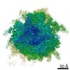
| Images |  1122.gif 1122.gif | 15.1 KB | ||
| Archive directory |  http://ftp.pdbj.org/pub/emdb/structures/EMD-1122ftp://ftp.pdbj.org/pub/emdb/structures/EMD-1122 http://ftp.pdbj.org/pub/emdb/structures/EMD-1122ftp://ftp.pdbj.org/pub/emdb/structures/EMD-1122 | HTTPS FTP |
-Validation report
| Summary document | emd_1122_validation.pdf.gz | 336.5 KB | Display | EMDB validaton report |
|---|---|---|---|---|
| Full document | emd_1122_full_validation.pdf.gz | 336.1 KB | Display | |
| Data in XML | emd_1122_validation.xml.gz | 5.9 KB | Display | |
| Arichive directory | https://ftp.pdbj.org/pub/emdb/validation_reports/EMD-1122ftp://ftp.pdbj.org/pub/emdb/validation_reports/EMD-1122 | HTTPS FTP |
-Related structure data
| Related structure data |  1zc8MC M: atomic model generated by this map C: citing same article ( |
|---|---|
| Similar structure data |
-Links
| EMDB pages | EMDB (EBI/PDBe) / EMDataResource |
|---|---|
| Related items in Molecule of the Month |


-Map
| File | Download / File: emd_1122.map.gz / Format: CCP4 / Size: 10.2 MB / Type: IMAGE STORED AS FLOATING POINT NUMBER (4 BYTES) | ||||||||||||||||||||||||||||||||||||||||||||||||||||||||||||||||||||
|---|---|---|---|---|---|---|---|---|---|---|---|---|---|---|---|---|---|---|---|---|---|---|---|---|---|---|---|---|---|---|---|---|---|---|---|---|---|---|---|---|---|---|---|---|---|---|---|---|---|---|---|---|---|---|---|---|---|---|---|---|---|---|---|---|---|---|---|---|---|
| Annotation | this is a ribosome consisting 70S + Psite tRNA + mRNA + EF-Tu.GDP + alanylated tmRNA + SmpB + Kirromycin + S1 | ||||||||||||||||||||||||||||||||||||||||||||||||||||||||||||||||||||
| Voxel size | X=Y=Z: 2.82 Å | ||||||||||||||||||||||||||||||||||||||||||||||||||||||||||||||||||||
| Density |
| ||||||||||||||||||||||||||||||||||||||||||||||||||||||||||||||||||||
| Symmetry | Space group: 1 | ||||||||||||||||||||||||||||||||||||||||||||||||||||||||||||||||||||
| Details | EMDB XML:
CCP4 map header:
| ||||||||||||||||||||||||||||||||||||||||||||||||||||||||||||||||||||
-Supplemental data
- Sample components
-Entire : 70S-Psite tRNA - mRNA - EF-Tu.GDP - alanylated tmRNA - SmpB - Kir...
| Entire | Name: 70S-Psite tRNA - mRNA - EF-Tu.GDP - alanylated tmRNA - SmpB - Kirromycin - S1 |
|---|---|
| Components |
|
-Supramolecule #1000: 70S-Psite tRNA - mRNA - EF-Tu.GDP - alanylated tmRNA - SmpB - Kir...
| Supramolecule | Name: 70S-Psite tRNA - mRNA - EF-Tu.GDP - alanylated tmRNA - SmpB - Kirromycin - S1 type: sample / ID: 1000 / Number unique components: 5 |
|---|
-Supramolecule #1: 70S Ribosome
| Supramolecule | Name: 70S Ribosome / type: complex / ID: 1 / Details: Thermus thermophilus / Recombinant expression: No / Ribosome-details: ribosome-prokaryote: ALL |
|---|---|
| Source (natural) | Organism: Thermus thermophilus HB8 (bacteria) |
-Macromolecule #1: tRNA
| Macromolecule | Name: tRNA / type: rna / ID: 1 / Details: P site / Classification: TRANSFER / Structure: SINGLE STRANDED / Synthetic?: No |
|---|---|
| Source (natural) | Organism: Thermus thermophilus HB8 (bacteria) |
-Macromolecule #2: EF-Tu
| Macromolecule | Name: EF-Tu / type: protein_or_peptide / ID: 2 / Recombinant expression: No |
|---|---|
| Source (natural) | Organism: Thermus thermophilus HB8 (bacteria) |
-Macromolecule #4: SmpB
| Macromolecule | Name: SmpB / type: protein_or_peptide / ID: 4 / Recombinant expression: No |
|---|---|
| Source (natural) | Organism: Thermus thermophilus HB8 (bacteria) |
-Macromolecule #3: tmRNA
| Macromolecule | Name: tmRNA / type: ligand / ID: 3 / Recombinant expression: No |
|---|---|
| Source (natural) | Organism: Thermus thermophilus HB8 (bacteria) |
-Experimental details
-Structure determination
| Method | cryo EM |
|---|---|
 Processing Processing | single particle reconstruction |
| Aggregation state | particle |
-Sample preparation
| Buffer | pH: 7.5 Details: 25 mM Hepes-KOH pH 7.5, 30 mM NH4Cl, 7mM MgCl2, 2 mM ATP, 6 mM PEP, 10g/ml PK |
|---|---|
| Vitrification | Cryogen name: ETHANE / Chamber humidity: 90 % / Chamber temperature: 93 K / Instrument: HOMEMADE PLUNGER Details: Vitrification instrument: two side blotting plunger Method: Blot for 2 seconds before plunging |
- Electron microscopy
| Microscope | FEI TECNAI F20 |
|---|---|
| Temperature | Average: 93 K |
| Date | Aug 1, 2002 |
| Image recording | Category: FILM / Film or detector model: KODAK SO-163 FILM / Digitization - Scanner: ZEISS SCAI / Digitization - Sampling interval: 14 µm / Number real images: 44 / Average electron dose: 15 e/Å2 / Od range: 1.2 / Bits/pixel: 12 |
| Electron beam | Acceleration voltage: 200 kV / Electron source:  FIELD EMISSION GUN FIELD EMISSION GUN |
| Electron optics | Calibrated magnification: 52000 / Illumination mode: FLOOD BEAM / Imaging mode: BRIGHT FIELD / Cs: 2.0 mm / Nominal defocus max: 3.7 µm / Nominal defocus min: 1.45 µm / Nominal magnification: 49000 |
| Sample stage | Specimen holder: Cryo transfer / Specimen holder model: GATAN LIQUID NITROGEN |
| Experimental equipment |  Model: Tecnai F20 / Image courtesy: FEI Company |
-Image processing
| CTF correction | Details: defocus groups |
|---|---|
| Final reconstruction | Applied symmetry - Point group: C1 (asymmetric) / Algorithm: OTHER / Resolution.type: BY AUTHOR / Resolution: 13.0 Å / Resolution method: OTHER / Software - Name: SPIDER / Number images used: 27644 |
| Final angle assignment | Details: SPIDER definition |
-Atomic model buiding 1
| Software | Name: manual |
|---|---|
| Details | Protocol: Rigid Body. The domains were separately fitted by manual docking using program O |
| Refinement | Protocol: RIGID BODY FIT / Target criteria: cross correlation coefficient |
| Output model | PDB-1zc8: |
