 Movie
Movie Controller

[English] 日本語
 Yorodumi
- EMDB-1110: Structural basis for the function of the ribosomal L7/12 stalk in... -
+ Open data

- Basic information
| Entry | Database: EMDB / ID: EMD-1110 | |||||||||
|---|---|---|---|---|---|---|---|---|---|---|
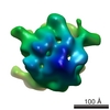
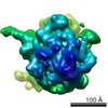
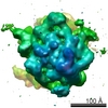
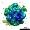
| Title | Structural basis for the function of the ribosomal L7/12 stalk in factor binding and GTPase activation. | |||||||||
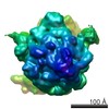
 Map data Map data | This is a map of the E.coli Ribosome in complex with elongation factor G in the presence of the antibiotic fusidic acid. | |||||||||
 Sample Sample |
| |||||||||
| Biological species |   Escherichia coli (E. coli) Escherichia coli (E. coli) | |||||||||
| Method | single particle reconstruction / cryo EM / Resolution: 18.0 Å | |||||||||
 Authors Authors | Diaconu M / Kothe U / Schlunzen F / Harms JM / Fischer N / Stark H / Rodnina MV / Wahl MC | |||||||||
 Citation Citation | Journal: Cell / Year: 2005 Title: Structural basis for the function of the ribosomal L7/12 stalk in factor binding and GTPase activation. Authors: Mihaela Diaconu / Ute Kothe / Frank Schlünzen / Niels Fischer / Jörg M Harms / Alexander G Tonevitsky / Holger Stark / Marina V Rodnina / Markus C Wahl /  Abstract: The L7/12 stalk of the large subunit of bacterial ribosomes encompasses protein L10 and multiple copies of L7/12. We present crystal structures of Thermotoga maritima L10 in complex with three L7/12 ...The L7/12 stalk of the large subunit of bacterial ribosomes encompasses protein L10 and multiple copies of L7/12. We present crystal structures of Thermotoga maritima L10 in complex with three L7/12 N-terminal-domain dimers, refine the structure of an archaeal L10E N-terminal domain on the 50S subunit, and identify these elements in cryo-electron-microscopic reconstructions of Escherichia coli ribosomes. The mobile C-terminal helix alpha8 of L10 carries three L7/12 dimers in T. maritima and two in E. coli, in concordance with the different length of helix alpha8 of L10 in these organisms. The stalk is organized into three elements (stalk base, L10 helix alpha8-L7/12 N-terminal-domain complex, and L7/12 C-terminal domains) linked by flexible connections. Highly mobile L7/12 C-terminal domains promote recruitment of translation factors to the ribosome and stimulate GTP hydrolysis by the ribosome bound factors through stabilization of their active GTPase conformation. | |||||||||
| History |
|
- Structure visualization
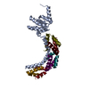
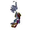
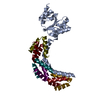
| Movie |
 Movie viewer Movie viewer |
|---|---|
| Structure viewer | EM map: SurfViewMolmilJmol/JSmol |
| Supplemental images |

- Downloads & links
-EMDB archive
| Map data | emd_1110.map.gz | 4.5 MB | EMDB map data format | |
|---|---|---|---|---|
| Header (meta data) | emd-1110-v30.xmlemd-1110.xml | 9.6 KB 9.6 KB | Display Display | EMDB header |
| Images | 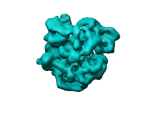 1110.gif 1110.gif | 46.5 KB | ||
| Archive directory |  http://ftp.pdbj.org/pub/emdb/structures/EMD-1110ftp://ftp.pdbj.org/pub/emdb/structures/EMD-1110 http://ftp.pdbj.org/pub/emdb/structures/EMD-1110ftp://ftp.pdbj.org/pub/emdb/structures/EMD-1110 | HTTPS FTP |
-Related structure data


-Links
| EMDB pages | EMDB (EBI/PDBe) / EMDataResource |
|---|---|
| Related items in Molecule of the Month |

-Map
| File | Download / File: emd_1110.map.gz / Format: CCP4 / Size: 4.7 MB / Type: IMAGE STORED AS FLOATING POINT NUMBER (4 BYTES) | ||||||||||||||||||||||||||||||||||||||||||||||||||||||||||||||||||||
|---|---|---|---|---|---|---|---|---|---|---|---|---|---|---|---|---|---|---|---|---|---|---|---|---|---|---|---|---|---|---|---|---|---|---|---|---|---|---|---|---|---|---|---|---|---|---|---|---|---|---|---|---|---|---|---|---|---|---|---|---|---|---|---|---|---|---|---|---|---|
| Annotation | This is a map of the E.coli Ribosome in complex with elongation factor G in the presence of the antibiotic fusidic acid. | ||||||||||||||||||||||||||||||||||||||||||||||||||||||||||||||||||||
| Voxel size | X=Y=Z: 3.2 Å | ||||||||||||||||||||||||||||||||||||||||||||||||||||||||||||||||||||
| Density |
| ||||||||||||||||||||||||||||||||||||||||||||||||||||||||||||||||||||
| Symmetry | Space group: 1 | ||||||||||||||||||||||||||||||||||||||||||||||||||||||||||||||||||||
| Details | EMDB XML:
CCP4 map header:
| ||||||||||||||||||||||||||||||||||||||||||||||||||||||||||||||||||||
-Supplemental data
- Sample components
-Entire : E coli ribosome in complex with elongation factor G
| Entire | Name: E coli ribosome in complex with elongation factor G |
|---|---|
| Components |
|
-Supramolecule #1000: E coli ribosome in complex with elongation factor G
| Supramolecule | Name: E coli ribosome in complex with elongation factor G / type: sample / ID: 1000 / Number unique components: 2 |
|---|---|
| Molecular weight | Experimental: 2.5 MDa |
-Supramolecule #1: 70S
| Supramolecule | Name: 70S / type: complex / ID: 1 / Recombinant expression: No / Ribosome-details: ribosome-prokaryote: ALL |
|---|---|
| Source (natural) | Organism: Escherichia coli (E. coli) |
| Molecular weight | Theoretical: 2.5 MDa |
-Macromolecule #1: Elongation Factor G
| Macromolecule | Name: Elongation Factor G / type: protein_or_peptide / ID: 1 / Recombinant expression: No |
|---|---|
| Source (natural) | Organism: Escherichia coli (E. coli) |
-Experimental details
-Structure determination
| Method | cryo EM |
|---|---|
 Processing Processing | single particle reconstruction |
| Aggregation state | particle |
-Sample preparation
| Concentration | 4.0 mg/mL |
|---|---|
| Buffer | pH: 7.5 Details: 50 mM Tris HCl, 70 mM NH4Cl, 30 mM KCl, 7 mM MgCl2, 1 mM DTT |
| Vitrification | Cryogen name: ETHANE / Chamber humidity: 40 % / Chamber temperature: 93 K / Instrument: HOMEMADE PLUNGER / Details: Vitrification instrument: home made / Method: Blotted manually for two seconds before plunging |
- Electron microscopy
| Microscope | FEI/PHILIPS CM200FEG |
|---|---|
| Electron beam | Acceleration voltage: 200 kV / Electron source: FIELD EMISSION GUN |
| Electron optics | Illumination mode: FLOOD BEAM / Imaging mode: BRIGHT FIELDBright-field microscopy / Cs: 2 mm / Nominal defocus max: 2.0 µm / Nominal defocus min: 1.2 µm |
| Sample stage | Specimen holder: Eucentric / Specimen holder model: GATAN LIQUID NITROGEN |
| Temperature | Average: 93 K |
| Image recording | Category: FILM / Film or detector model: KODAK SO-163 FILM / Digitization - Scanner: PRIMESCAN / Digitization - Sampling interval: 4.0 µm / Number real images: 12 / Average electron dose: 15 e/Å2 / Od range: 1.3 / Bits/pixel: 8 |
-Image processing
| CTF correction | Details: Each particle |
|---|---|
| Final reconstruction | Applied symmetry - Point group: C1 (asymmetric) / Algorithm: OTHER / Resolution.type: BY AUTHOR / Resolution: 18.0 Å / Resolution method: OTHER / Software - Name: IMAGIC / Number images used: 8000 |
| Details | The particles were selected manually. |
-Atomic model buiding 1
| Software | Name: Amira 3.1, TGS Software |
|---|---|
| Details | Protocol: Rigid Body. Manual fitting |
| Refinement | Protocol: RIGID BODY FIT |
