 Movie
Movie Controller

[English] 日本語
 Yorodumi
- EMDB-1068: Three-dimensional structures of translating ribosomes by Cryo-EM. -
+ Open data

- Basic information
| Entry | Database: EMDB / ID: EMD-1068 | |||||||||
|---|---|---|---|---|---|---|---|---|---|---|
| Title | Three-dimensional structures of translating ribosomes by Cryo-EM. | |||||||||
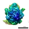
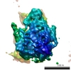
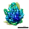
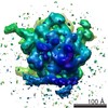
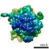
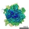
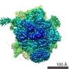
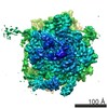
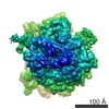
 Map data Map data | 3D reconstruction of inactive 70S E. coli ribosome | |||||||||
 Sample Sample |
| |||||||||
| Biological species |   Escherichia coli (E. coli) Escherichia coli (E. coli) | |||||||||
| Method | single particle reconstruction / cryo EM / Resolution: 13.4 Å | |||||||||
 Authors Authors | Gilbert RJC / Fucini P / Connell S / Fuller SD / Nierhaus KH / Robinson CV / Dobson CM / Stuart DI | |||||||||
 Citation Citation | Journal: Mol Cell / Year: 2004 Title: Three-dimensional structures of translating ribosomes by Cryo-EM. Authors: Robert J C Gilbert / Paola Fucini / Sean Connell / Stephen D Fuller / Knud H Nierhaus / Carol V Robinson / Christopher M Dobson / David I Stuart /  Abstract: Cryo-electron microscopy and image reconstruction techniques have been used to obtain three-dimensional maps for E. coli ribosomes stalled following translation of three representative proteins. ...Cryo-electron microscopy and image reconstruction techniques have been used to obtain three-dimensional maps for E. coli ribosomes stalled following translation of three representative proteins. Comparisons of these electron density maps, at resolutions of between 13 and 16 A, with that of a nontranslating ribosome pinpoint specific structural differences in stalled ribosomes and identify additional material, including tRNAs and mRNA. In addition, the tunnel through the large subunit, the anticipated exit route of newly synthesized proteins, is partially occluded in all the stalled ribosome structures. This observation suggests that significant segments of the nascent polypeptide chains examined here could be located within an expanded tunnel, perhaps in a rudimentary globular conformation. Such behavior could be an important aspect of the folding of at least some proteins in the cellular environment. | |||||||||
| History |
|
- Structure visualization
| Movie |
 Movie viewer Movie viewer |
|---|---|
| Structure viewer | EM map: SurfViewMolmilJmol/JSmol |
| Supplemental images |
- Downloads & links
-EMDB archive
| Map data | emd_1068.map.gz | 7.4 MB | EMDB map data format | |
|---|---|---|---|---|
| Header (meta data) | emd-1068-v30.xmlemd-1068.xml | 10 KB 10 KB | Display Display | EMDB header |
| Images |  1068.gif 1068.gif | 84.2 KB | ||
| Archive directory |  http://ftp.pdbj.org/pub/emdb/structures/EMD-1068ftp://ftp.pdbj.org/pub/emdb/structures/EMD-1068 http://ftp.pdbj.org/pub/emdb/structures/EMD-1068ftp://ftp.pdbj.org/pub/emdb/structures/EMD-1068 | HTTPS FTP |
-Related structure data


-Links
| EMDB pages | EMDB (EBI/PDBe) / EMDataResource |
|---|---|
| Related items in Molecule of the Month |

-Map
| File | Download / File: emd_1068.map.gz / Format: CCP4 / Size: 7.8 MB / Type: IMAGE STORED AS FLOATING POINT NUMBER (4 BYTES) | ||||||||||||||||||||||||||||||||||||||||||||||||||||||||||||||||||||
|---|---|---|---|---|---|---|---|---|---|---|---|---|---|---|---|---|---|---|---|---|---|---|---|---|---|---|---|---|---|---|---|---|---|---|---|---|---|---|---|---|---|---|---|---|---|---|---|---|---|---|---|---|---|---|---|---|---|---|---|---|---|---|---|---|---|---|---|---|---|
| Annotation | 3D reconstruction of inactive 70S E. coli ribosome | ||||||||||||||||||||||||||||||||||||||||||||||||||||||||||||||||||||
| Voxel size | X=Y=Z: 3.33 Å | ||||||||||||||||||||||||||||||||||||||||||||||||||||||||||||||||||||
| Density |
| ||||||||||||||||||||||||||||||||||||||||||||||||||||||||||||||||||||
| Symmetry | Space group: 1 | ||||||||||||||||||||||||||||||||||||||||||||||||||||||||||||||||||||
| Details | EMDB XML:
CCP4 map header:
| ||||||||||||||||||||||||||||||||||||||||||||||||||||||||||||||||||||
-Supplemental data
- Sample components
-Entire : 70S E. coli ribosome
| Entire | Name: 70S E. coli ribosome |
|---|---|
| Components |
|
-Supramolecule #1000: 70S E. coli ribosome
| Supramolecule | Name: 70S E. coli ribosome / type: sample / ID: 1000 / Details: The sample was monodisperse. / Oligomeric state: 30S subunit and 50S subunit / Number unique components: 2 |
|---|---|
| Molecular weight | Experimental: 2 MDa |
-Supramolecule #1: E. coli 30S subunit
| Supramolecule | Name: E. coli 30S subunit / type: complex / ID: 1 / Name.synonym: 30S / Recombinant expression: No / Ribosome-details: ribosome-prokaryote: ALL |
|---|---|
| Source (natural) | Organism: Escherichia coli (E. coli) |
-Supramolecule #2: E. coli 50S subunit
| Supramolecule | Name: E. coli 50S subunit / type: complex / ID: 2 / Name.synonym: 50S / Recombinant expression: No / Ribosome-details: ribosome-prokaryote: ALL |
|---|---|
| Source (natural) | Organism: Escherichia coli (E. coli) |
-Experimental details
-Structure determination
| Method | cryo EM |
|---|---|
 Processing Processing | single particle reconstruction |
| Aggregation state | particle |
-Sample preparation
| Concentration | 5.0 mg/mL |
|---|---|
| Buffer | Details: 20 mM HEPES, 150 mM ammonium acetate, 6 mM magnesium acetate, 2 mM spermidine, 0.05 mM spermine and 4 mM 2-mercaptoethanol. Concentration of ribosomes expressed to A260 units. |
| Grid | Details: 300 mesh copper grid with holey carbon film |
| Vitrification | Cryogen name: ETHANE / Chamber temperature: 100 K / Instrument: HOMEMADE PLUNGER Details: Vitrification instrument: Standard unmodified guillotine plunger |
- Electron microscopy
| Microscope | FEI/PHILIPS CM200FEG |
|---|---|
| Electron beam | Acceleration voltage: 200 kV / Electron source: FIELD EMISSION GUN |
| Electron optics | Illumination mode: OTHER / Imaging mode: OTHER / Cs: 2 mm / Nominal defocus max: 3.915 µm / Nominal defocus min: 1.475 µm / Nominal magnification: 50000 |
| Sample stage | Specimen holder: Eucentric / Specimen holder model: GATAN LIQUID NITROGEN |
| Temperature | Average: 100 K |
| Date | May 1, 2000 |
| Image recording | Category: FILM / Film or detector model: KODAK SO-163 FILM / Digitization - Scanner: ZEISS SCAI / Digitization - Sampling interval: 7 µm / Number real images: 12 / Od range: 5 / Bits/pixel: 8 |
-Image processing
| CTF correction | Details: Each negative dataset |
|---|---|
| Final angle assignment | Details: SPIDER euler |
| Final reconstruction | Applied symmetry - Point group: C1 (asymmetric) / Algorithm: OTHER / Resolution.type: BY AUTHOR / Resolution: 13.4 Å / Resolution method: FSC 0.5 CUT-OFF / Software - Name: SPIDER, IMAGIC, GAP, CNS, XPLOR Details: Final maps were calculated from 12 individual datasets, with scaling in reciprocal space to crystallographic ribosome structures and correction for map anisotropy by B-factor weighting of amplitudes in XPLOR. Number images used: 8238 |
-Atomic model buiding 1
| Software | Name: GAP |
|---|---|
| Details | Protocol: Rigid body |
| Refinement | Space: REAL / Protocol: RIGID BODY FIT / Target criteria: R-factor and correlation coefficient |
