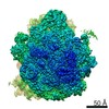
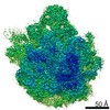
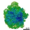
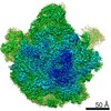
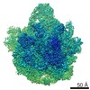
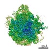
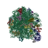
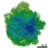
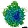
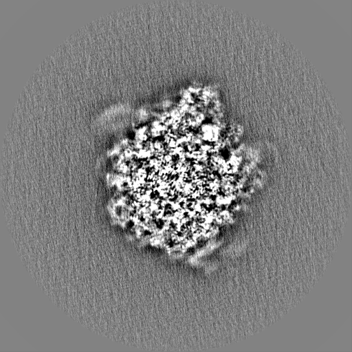
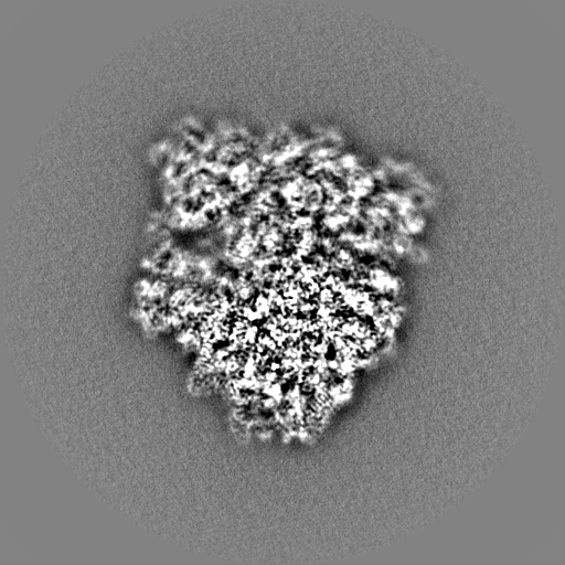
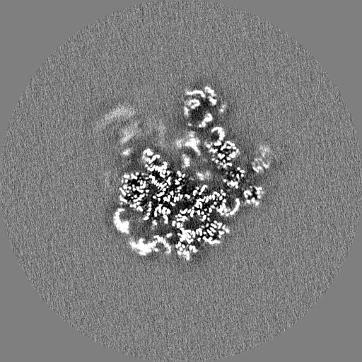
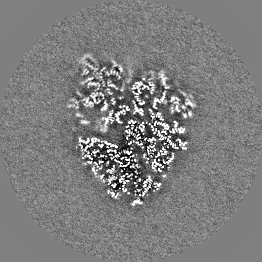
- EMDB-10655: E. coli 50S ribosomal subunit in complex with dirithromycin, fMet... -
+
データを開く
IDまたはキーワード:
読み込み中...
-
基本情報
登録情報
データベース: EMDB / ID: EMD-10655
タイトル
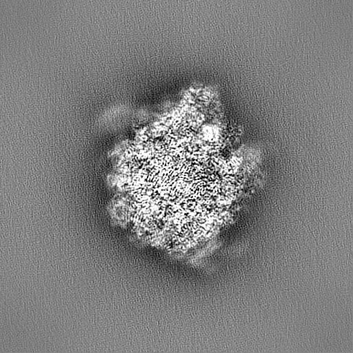
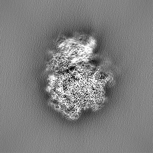
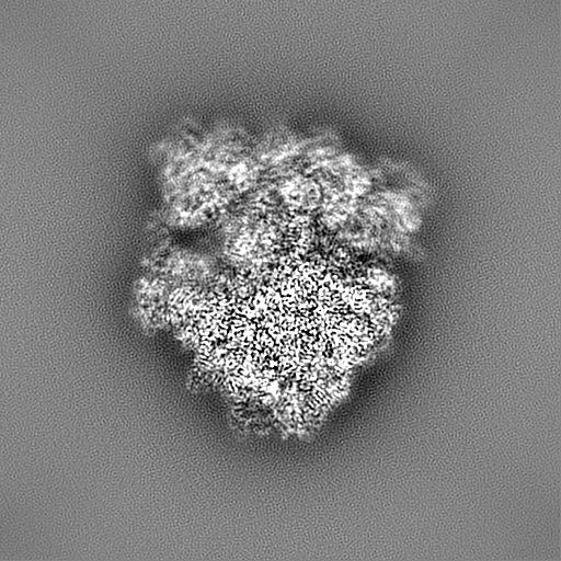
E. coli 50S ribosomal subunit in complex with dirithromycin, fMet-Phe-tRNA(Phe) and deacylated tRNA(iMet).
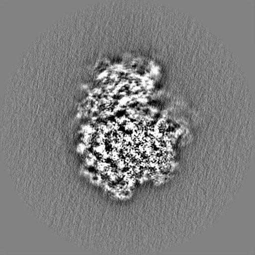
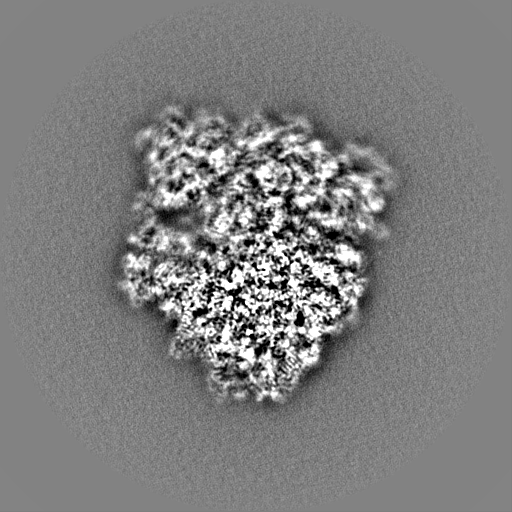
マップデータ
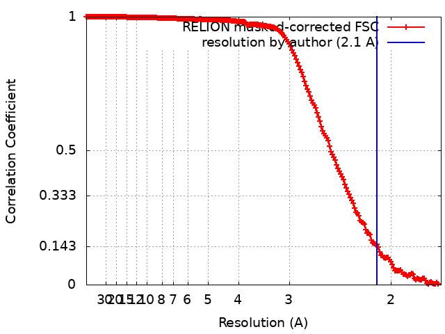
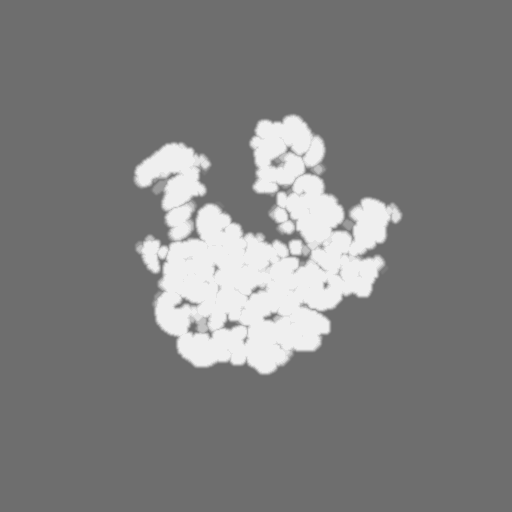
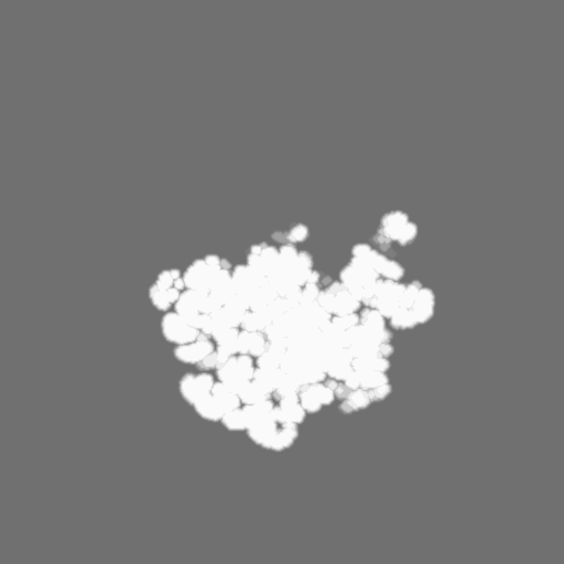
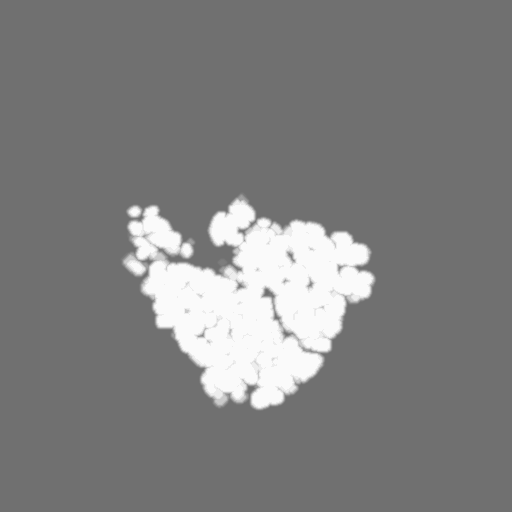
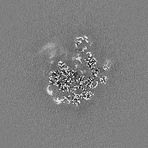
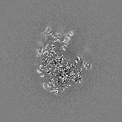
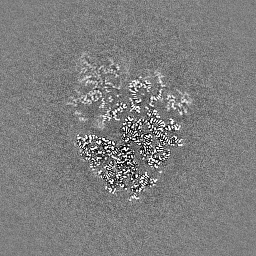
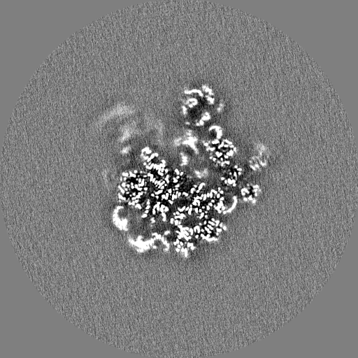
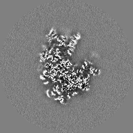
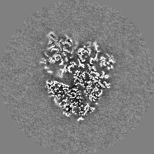
Sharpened map of the 50S LSU with Dirithromycin after high-order aberration correction in Relion 3.1
試料
複合体: E. coli 50S ribosomal subunit in complex with dirithromycin, fMet-Phe-tRNA(Phe) and deacylated tRNA(iMet).
複合体: E. coli 50S ribosomal subunit
RNA: x 3種
タンパク質・ペプチド: x 29種
複合体: fMet-Phe-tRNA(Phe)
RNA: x 1種
リガンド: x 3種
機能・相同性
機能・相同性情報
stringent response / transcriptional attenuation / endoribonuclease inhibitor activity / RNA-binding transcription regulator activity / positive regulation of ribosome biogenesis / negative regulation of cytoplasmic translation / translational termination / DnaA-L2 complex / translation repressor activity / negative regulation of DNA-templated DNA replication initiation ...stringent response / transcriptional attenuation / endoribonuclease inhibitor activity / RNA-binding transcription regulator activity / positive regulation of ribosome biogenesis / negative regulation of cytoplasmic translation / translational termination / DnaA-L2 complex / translation repressor activity / negative regulation of DNA-templated DNA replication initiation / ribosome assembly / mRNA regulatory element binding translation repressor activity / assembly of large subunit precursor of preribosome / cytosolic ribosome assembly / response to reactive oxygen species / regulation of cell growth / DNA-templated transcription termination / response to radiation / ribosomal large subunit assembly / mRNA 5'-UTR binding / large ribosomal subunit / ribosome binding / 5S rRNA binding / large ribosomal subunit rRNA binding / transferase activity / cytosolic large ribosomal subunit / cytoplasmic translation / tRNA binding / negative regulation of translation / rRNA binding / リボソーム / structural constituent of ribosome / 翻訳 (生物学) / response to antibiotic / negative regulation of DNA-templated transcription / mRNA binding / DNA binding / RNA binding / zinc ion binding / 細胞質基質 / 細胞質 類似検索 - 分子機能
Ribosomal protein L10, eubacterial, conserved site / Ribosomal protein L10 signature. / Ribosomal protein L10 / : / Ribosomal protein L25, short-form / Ribosomal protein L11, bacterial-type / Ribosomal protein L21, conserved site / Ribosomal protein L11, conserved site / Ribosomal protein L11 signature. / Ribosomal protein L21 signature. ...Ribosomal protein L10, eubacterial, conserved site / Ribosomal protein L10 signature. / Ribosomal protein L10 / : / Ribosomal protein L25, short-form / Ribosomal protein L11, bacterial-type / Ribosomal protein L21, conserved site / Ribosomal protein L11, conserved site / Ribosomal protein L11 signature. / Ribosomal protein L21 signature. / Ribosomal protein L10-like domain superfamily / Ribosomal protein L10 / Ribosomal protein L10P / Ribosomal protein L16 signature 1. / : / Ribosomal protein L6, conserved site / Ribosomal protein L6 signature 1. / Ribosomal protein L16, conserved site / Ribosomal protein L16 signature 2. / Ribosomal protein L17 signature. / Ribosomal L25p family / Ribosomal protein L25 / Ribosomal protein L11, N-terminal / Ribosomal protein L11, N-terminal domain / Ribosomal protein L11/L12 / Ribosomal protein L11, C-terminal / Ribosomal protein L11, C-terminal domain superfamily / Ribosomal protein L11/L12, N-terminal domain superfamily / Ribosomal protein L11, RNA binding domain / Ribosomal protein L11/L12 / Ribosomal protein L28/L24 superfamily / Ribosomal protein L36 signature. / Ribosomal protein L25/Gln-tRNA synthetase, N-terminal / Ribosomal protein L25/Gln-tRNA synthetase, anti-codon-binding domain superfamily / Ribosomal protein L32p, bacterial type / Ribosomal protein L28 / Ribosomal protein L35, conserved site / Ribosomal protein L35 signature. / Ribosomal protein L33, conserved site / Ribosomal protein L33 signature. / Ribosomal protein L35, non-mitochondrial / Ribosomal protein L5, bacterial-type / Ribosomal protein L6, bacterial-type / Ribosomal protein L18, bacterial-type / Ribosomal protein L19, conserved site / Ribosomal protein L19 signature. / Ribosomal protein L36 / Ribosomal protein L36 superfamily / Ribosomal protein L36 / Ribosomal protein L20 signature. / Ribosomal protein L27, conserved site / Ribosomal protein L27 signature. / Ribosomal protein L14P, bacterial-type / Ribosomal protein L34, conserved site / Ribosomal protein L34 signature. / Ribosomal protein L22, bacterial/chloroplast-type / Ribosomal protein L35 / Ribosomal protein L35 superfamily / Ribosomal protein L2, bacterial/organellar-type / Ribosomal protein L35 / Ribosomal L28 family / Ribosomal protein L33 / Ribosomal protein L33 / Ribosomal protein L28/L24 / Ribosomal protein L33 superfamily / Ribosomal protein L30, bacterial-type / : / Ribosomal protein L16 / Ribosomal protein L18 / Ribosomal L18 of archaea, bacteria, mitoch. and chloroplast / L28p-like / Ribosomal protein L20 / Ribosomal protein L20 / Ribosomal protein L20, C-terminal / Ribosomal protein L21 / Ribosomal protein L27 / Ribosomal L27 protein / Ribosomal protein L19 / Ribosomal protein L19 superfamily / Ribosomal protein L19 / Ribosomal proteins 50S L24/mitochondrial 39S L24 / Ribosomal protein L17 / Ribosomal protein L17 superfamily / Ribosomal protein L17 / Ribosomal protein L21-like / L21-like superfamily / Ribosomal prokaryotic L21 protein / Ribosomal L32p protein family / Ribosomal protein L24 / Ribosomal protein L32p / Ribosomal protein L34 / Ribosomal protein L34 / Ribosomal protein L13, bacterial-type / Ribosomal protein L3, bacterial/organelle-type / Ribosomal protein L23/L25, conserved site / Ribosomal protein L15, bacterial-type / 50S ribosomal protein uL4 / Ribosomal protein L23 signature. / Ribosomal protein L30, conserved site / Ribosomal protein L30 signature. 類似検索 - ドメイン・相同性
Large ribosomal subunit protein uL15 / Large ribosomal subunit protein uL10 / Large ribosomal subunit protein uL11 / Large ribosomal subunit protein bL19 / Large ribosomal subunit protein bL20 / Large ribosomal subunit protein bL27 / Large ribosomal subunit protein bL28 / Large ribosomal subunit protein uL29 / Large ribosomal subunit protein bL32 / Large ribosomal subunit protein bL33 ...Large ribosomal subunit protein uL15 / Large ribosomal subunit protein uL10 / Large ribosomal subunit protein uL11 / Large ribosomal subunit protein bL19 / Large ribosomal subunit protein bL20 / Large ribosomal subunit protein bL27 / Large ribosomal subunit protein bL28 / Large ribosomal subunit protein uL29 / Large ribosomal subunit protein bL32 / Large ribosomal subunit protein bL33 / Large ribosomal subunit protein bL34 / Large ribosomal subunit protein bL35 / Large ribosomal subunit protein bL36A / Large ribosomal subunit protein uL13 / Large ribosomal subunit protein uL14 / Large ribosomal subunit protein uL16 / Large ribosomal subunit protein uL23 / Large ribosomal subunit protein bL17 / Large ribosomal subunit protein bL21 / Large ribosomal subunit protein uL30 / Large ribosomal subunit protein uL6 / Large ribosomal subunit protein uL18 / Large ribosomal subunit protein uL2 / Large ribosomal subunit protein uL3 / Large ribosomal subunit protein uL24 / Large ribosomal subunit protein uL4 / Large ribosomal subunit protein uL22 / Large ribosomal subunit protein uL5 / Large ribosomal subunit protein bL25 類似検索 - 構成要素
National Institutes of Health/National Institute Of Allergy and Infectious Diseases (NIH/NIAID)
R21-AI137584
米国
National Institutes of Health/National Institute of General Medical Sciences (NIH/NIGMS)
R01-GM132302
米国
Russian Science Foundation
18-44-04005
ロシア
Russian Foundation for Basic Research
17-00-00366
ロシア
引用
ジャーナル: RNA / 年: 2020 タイトル: Insights into the improved macrolide inhibitory activity from the high-resolution cryo-EM structure of dirithromycin bound to the 70S ribosome. 著者: Evgeny B Pichkur / Alena Paleskava / Andrey G Tereshchenkov / Pavel Kasatsky / Ekaterina S Komarova / Dmitrii I Shiriaev / Alexey A Bogdanov / Olga A Dontsova / Ilya A Osterman / Petr V ...著者: Evgeny B Pichkur / Alena Paleskava / Andrey G Tereshchenkov / Pavel Kasatsky / Ekaterina S Komarova / Dmitrii I Shiriaev / Alexey A Bogdanov / Olga A Dontsova / Ilya A Osterman / Petr V Sergiev / Yury S Polikanov / Alexander G Myasnikov / Andrey L Konevega / 要旨: Macrolides are one of the most successful and widely used classes of antibacterials, which kill or stop the growth of pathogenic bacteria by binding near the active site of the ribosome and ...Macrolides are one of the most successful and widely used classes of antibacterials, which kill or stop the growth of pathogenic bacteria by binding near the active site of the ribosome and interfering with protein synthesis. Dirithromycin is a derivative of the prototype macrolide erythromycin with additional hydrophobic side chain. In our recent study, we have discovered that the side chain of dirithromycin forms lone pair-π stacking interaction with the aromatic imidazole ring of the His69 residue in ribosomal protein uL4 of the 70S ribosome. In the current work, we found that neither the presence of the side chain, nor the additional contact with the ribosome, improve the binding affinity of dirithromycin to the ribosome. Nevertheless, we found that dirithromycin is a more potent inhibitor of in vitro protein synthesis in comparison with its parent compound, erythromycin. Using high-resolution cryo-electron microscopy, we determined the structure of the dirithromycin bound to the translating 70S ribosome, which suggests that the better inhibitory properties of the drug could be rationalized by the side chain of dirithromycin pointing into the lumen of the nascent peptide exit tunnel, where it can interfere with the normal passage of the growing polypeptide chain.
 ムービー
ムービー コントローラー


 データを開く
 基本情報
基本情報 マップデータ
 試料
試料 機能・相同性情報
 stringent response /

 データ登録者
 ロシア,
ロシア,  米国, 5件
  引用
引用
 構造の表示
構造の表示
 ダウンロードとリンク
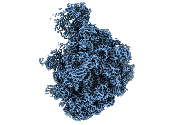 emd_10655.png
 http://ftp.pdbj.org/pub/emdb/structures/EMD-10655


 Z
Z Y
Y X
X


 試料の構成要素

 解析
解析 電子顕微鏡法