 Movie
Movie Controller

[English] 日本語
 Yorodumi
- EMDB-1022: Three-dimensional reconstructions from cryoelectron microscopy im... -
+ Open data

- Basic information
| Entry | Database: EMDB / ID: EMD-1022 | |||||||||
|---|---|---|---|---|---|---|---|---|---|---|
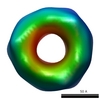
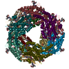
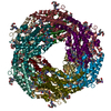
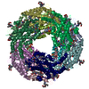
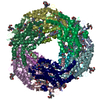
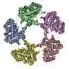
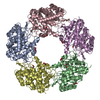
| Title | Three-dimensional reconstructions from cryoelectron microscopy images reveal an intimate complex between helicase DnaB and its loading partner DnaC. | |||||||||
 Map data Map data | DnaB | |||||||||
 Sample Sample |
| |||||||||
| Function / homology | : / DNA helicase activity Function and homology information Function and homology information | |||||||||
| Biological species |  | |||||||||
| Method | single particle reconstruction / cryo EM / Resolution: 34.5 Å | |||||||||
 Authors Authors | San Martin C | |||||||||
 Citation Citation | Journal: Structure / Year: 1998 Title: Three-dimensional reconstructions from cryoelectron microscopy images reveal an intimate complex between helicase DnaB and its loading partner DnaC. Authors: C San Martin / M Radermacher / B Wolpensinger / A Engel / C S Miles / N E Dixon / J M Carazo /  Abstract: BACKGROUND: DNA helicases play a fundamental role in all aspects of nucleic acid metabolism and defects in these enzymes have been implicated in a number of inherited human disorders. DnaB is the ...BACKGROUND: DNA helicases play a fundamental role in all aspects of nucleic acid metabolism and defects in these enzymes have been implicated in a number of inherited human disorders. DnaB is the major replicative DNA helicase in Escherichia coli and has been used as a model system for studying the structure and function of hexameric helicases. The native protein is a hexamer of identical subunits, which in solution forms a complex with six molecules of the loading protein DnaC. DnaB is delivered from this complex onto the DNA template, with the subsequent release of DnaC. We report here the structures of the DnaB helicase hexamer and its complex with DnaC under a defined set of experimental conditions, as determined by three-dimensional cryoelectron microscopy. It was hoped that the structures would provide insight into the mechanisms of helicase activity. RESULTS: The DnaB structure reveals that six DnaB monomers assemble as three asymmetric dimers to form a polar, ring-like hexamer. The hexamer has two faces, one displaying threefold and the other ...RESULTS: The DnaB structure reveals that six DnaB monomers assemble as three asymmetric dimers to form a polar, ring-like hexamer. The hexamer has two faces, one displaying threefold and the other sixfold symmetry. The six DnaC protomers bind tightly to the sixfold face of the DnaB hexamer. This is the first report of a three-dimensional structure of a helicase obtained using cryoelectron microscopy, and the first report of the structure of a helicase in complex with a loading protein. CONCLUSIONS: The structures of the DnaB helicase and its complex with DnaC reveal some interesting structural features relevant to helicase function and to the assembly of the two-protein complex. ...CONCLUSIONS: The structures of the DnaB helicase and its complex with DnaC reveal some interesting structural features relevant to helicase function and to the assembly of the two-protein complex. The results presented here provide a basis for a more complete understanding of the structure and function of these important proteins. | |||||||||
| History |
|
- Structure visualization
| Movie |
Movie viewer |
|---|---|
| Structure viewer | EM map: SurfViewMolmilJmol/JSmol |
| Supplemental images |
 UCSF Chimera
- Downloads & links
-EMDB archive
| Map data | emd_1022.map.gz | 451.7 KB | EMDB map data format | |
|---|---|---|---|---|
| Header (meta data) | emd-1022-v30.xmlemd-1022.xml | 8.6 KB 8.6 KB | Display Display | EMDB header |
| Images |  1022.gif 1022.gif | 12.9 KB | ||
| Archive directory |  http://ftp.pdbj.org/pub/emdb/structures/EMD-1022ftp://ftp.pdbj.org/pub/emdb/structures/EMD-1022 http://ftp.pdbj.org/pub/emdb/structures/EMD-1022ftp://ftp.pdbj.org/pub/emdb/structures/EMD-1022 | HTTPS FTP |
-Validation report
| Summary document | emd_1022_validation.pdf.gz | 211.8 KB | Display | EMDB validaton report |
|---|---|---|---|---|
| Full document | emd_1022_full_validation.pdf.gz | 210.9 KB | Display | |
| Data in XML | emd_1022_validation.xml.gz | 4.9 KB | Display | |
| Arichive directory | https://ftp.pdbj.org/pub/emdb/validation_reports/EMD-1022ftp://ftp.pdbj.org/pub/emdb/validation_reports/EMD-1022 | HTTPS FTP |
-Related structure data


-Links
| EMDB pages | EMDB (EBI/PDBe) / EMDataResource |
|---|
-Map
| File | Download / File: emd_1022.map.gz / Format: CCP4 / Size: 478.5 KB / Type: IMAGE STORED AS FLOATING POINT NUMBER (4 BYTES) | ||||||||||||||||||||||||||||||||||||||||||||||||||||||||||||
|---|---|---|---|---|---|---|---|---|---|---|---|---|---|---|---|---|---|---|---|---|---|---|---|---|---|---|---|---|---|---|---|---|---|---|---|---|---|---|---|---|---|---|---|---|---|---|---|---|---|---|---|---|---|---|---|---|---|---|---|---|---|
| Annotation | DnaB | ||||||||||||||||||||||||||||||||||||||||||||||||||||||||||||
| Voxel size | X=Y=Z: 3.8 Å | ||||||||||||||||||||||||||||||||||||||||||||||||||||||||||||
| Density |
| ||||||||||||||||||||||||||||||||||||||||||||||||||||||||||||
| Symmetry | Space group: 1 | ||||||||||||||||||||||||||||||||||||||||||||||||||||||||||||
| Details | EMDB XML:
CCP4 map header:
| ||||||||||||||||||||||||||||||||||||||||||||||||||||||||||||
-Supplemental data
- Sample components
-Entire : DnaB from E.coli
| Entire | Name: DnaB from E.coli |
|---|---|
| Components |
|
-Supramolecule #1000: DnaB from E.coli
| Supramolecule | Name: DnaB from E.coli / type: sample / ID: 1000 / Oligomeric state: homohexamer / Number unique components: 1 |
|---|---|
| Molecular weight | Theoretical: 300 KDa |
-Macromolecule #1: DnaB helicase
| Macromolecule | Name: DnaB helicase / type: protein_or_peptide / ID: 1 / Name.synonym: DnaB / Number of copies: 6 / Oligomeric state: homohexamer / Recombinant expression: Yes |
|---|---|
| Source (natural) | Organism: |
| Molecular weight | Theoretical: 300 KDa |
| Recombinant expression | Organism: AN1459/pPS560 / Recombinant plasmid: pPS560 |
| Sequence | GO: DNA helicase activity / InterPro: INTERPRO: IPR001198 |
-Experimental details
-Structure determination
| Method | cryo EM |
|---|---|
 Processing Processing | single particle reconstruction |
| Aggregation state | particle |
-Sample preparation
| Concentration | .04 mg/mL |
|---|---|
| Buffer | pH: 7.6 Details: 50 mM Tris.HCl pH 7.6 2 mM DTT 5 mM MgCl2 200 mM NaCl 0.25 mM ADP |
| Vitrification | Cryogen name: ETHANE / Instrument: HOMEMADE PLUNGER Details: Vitrification instrument: double side blotting device Method: samples were adsorbed onto carbon-coated molybdenum holey grids after 30s glow discharge, and vitrified by quick plunging in liquid ethane in a double-side blotting device |
- Electron microscopy
| Microscope | FEI/PHILIPS EM420 |
|---|---|
| Temperature | Average: 98 K |
| Image recording | Category: FILM / Film or detector model: KODAK SO-163 FILM / Digitization - Scanner: EIKONIX IEEE 488 / Number real images: 27 / Average electron dose: 10 e/Å2 / Bits/pixel: 8 |
| Electron beam | Acceleration voltage: 100 kV / Electron source: LAB6 |
| Electron optics | Illumination mode: FLOOD BEAM / Imaging mode: BRIGHT FIELD / Cs: 2 mm / Nominal defocus max: 2.0 µm / Nominal defocus min: 1.5 µm / Nominal magnification: 60000 |
| Sample stage | Specimen holder: Gatan626 / Specimen holder model: GATAN LIQUID NITROGEN / Tilt angle max: 45 |
-Image processing
| Final reconstruction | Applied symmetry - Point group: C3 (3 fold cyclic) / Algorithm: OTHER / Resolution.type: BY AUTHOR / Resolution: 34.5 Å / Resolution method: OTHER / Software - Name: Xmipp, SPIDER / Number images used: 1369 |
|---|
