Movie
Movie Controller
 Structure viewers
 About Yorodumi Papers


+Search query
-Structure paper
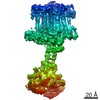
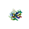
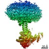
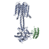
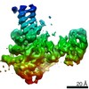
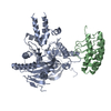
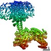
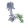
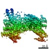
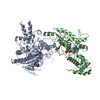
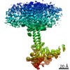
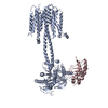
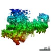
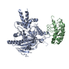
| Title | Structural basis of adenylyl cyclase 9 activation. |
|---|---|
| Journal, issue, pages | Nat Commun, Vol. 13, Issue 1, Page 1045, Year 2022 |
| Publish date | Feb 24, 2022 |
 Authors Authors | Chao Qi / Pia Lavriha / Ved Mehta / Basavraj Khanppnavar / Inayathulla Mohammed / Yong Li / Michalis Lazaratos / Jonas V Schaefer / Birgit Dreier / Andreas Plückthun / Ana-Nicoleta Bondar / Carmen W Dessauer / Volodymyr M Korkhov /    |
| PubMed Abstract | Adenylyl cyclase 9 (AC9) is a membrane-bound enzyme that converts ATP into cAMP. The enzyme is weakly activated by forskolin, fully activated by the G protein Gαs subunit and is autoinhibited by the ...Adenylyl cyclase 9 (AC9) is a membrane-bound enzyme that converts ATP into cAMP. The enzyme is weakly activated by forskolin, fully activated by the G protein Gαs subunit and is autoinhibited by the AC9 C-terminus. Although our recent structural studies of the AC9-Gαs complex provided the framework for understanding AC9 autoinhibition, the conformational changes that AC9 undergoes in response to activator binding remains poorly understood. Here, we present the cryo-EM structures of AC9 in several distinct states: (i) AC9 bound to a nucleotide inhibitor MANT-GTP, (ii) bound to an artificial activator (DARPin C4) and MANT-GTP, (iii) bound to DARPin C4 and a nucleotide analogue ATPαS, (iv) bound to Gαs and MANT-GTP. The artificial activator DARPin C4 partially activates AC9 by binding at a site that overlaps with the Gαs binding site. Together with the previously observed occluded and forskolin-bound conformations, structural comparisons of AC9 in the four conformations described here show that secondary structure rearrangements in the region surrounding the forskolin binding site are essential for AC9 activation. |
 External links External links | Nat Commun / PubMed:35210418 / PubMed Central |
| Methods | EM (single particle) |
| Resolution | 3.8 - 4.9 Å |
| Structure data | EMDB-13330, PDB-7pd4: EMDB-13331, PDB-7pd8: EMDB-13334, PDB-7pdd: EMDB-13335, PDB-7pde: EMDB-13336, PDB-7pdf: EMDB-13337, PDB-7pdg: EMDB-13338, PDB-7pdh: |
| Chemicals |  ChemComp-GSP:  ChemComp-MG: |
| Source |
|
 Keywords Keywords |  SIGNALING PROTEIN / membrane protein / adenylyl cyclase / signalling transduction. SIGNALING PROTEIN / membrane protein / adenylyl cyclase / signalling transduction. |