Movie
Movie Controller
 Structure viewers
 About Yorodumi Papers


+Search query
-Structure paper
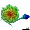
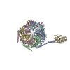
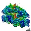
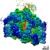
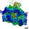
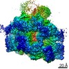
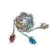
| Title | Structural basis for aggregate dissolution and refolding by the Mycobacterium tuberculosis ClpB-DnaK bi-chaperone system. |
|---|---|
| Journal, issue, pages | Cell Rep, Vol. 35, Issue 8, Page 109166, Year 2021 |
| Publish date | May 25, 2021 |
 Authors Authors | Yanting Yin / Xiang Feng / Hongjun Yu / Allison Fay / Amanda Kovach / Michael S Glickman / Huilin Li /  |
| PubMed Abstract | The M. tuberculosis (Mtb) ClpB is a protein disaggregase that helps to rejuvenate the bacterial cell. DnaK is a protein foldase that can function alone, but it can also bind to the ClpB hexamer to ...The M. tuberculosis (Mtb) ClpB is a protein disaggregase that helps to rejuvenate the bacterial cell. DnaK is a protein foldase that can function alone, but it can also bind to the ClpB hexamer to physically couple protein disaggregation with protein refolding, although the molecular mechanism is not well understood. Here, we report the cryo-EM analysis of the Mtb ClpB-DnaK bi-chaperone in the presence of ATPγS and a protein substrate. We observe three ClpB conformations in the presence of DnaK, identify a conserved TGIP loop linking the oligonucleotide/oligosaccharide-binding domain and the nucleotide-binding domain that is important for ClpB function, derive the interface between the regulatory middle domain of the ClpB and the DnaK nucleotide-binding domain, and find that DnaK binding stabilizes, but does not bend or tilt, the ClpB middle domain. We propose a model for the synergistic actions of aggregate dissolution and refolding by the Mtb ClpB-DnaK bi-chaperone system. |
 External links External links | Cell Rep / PubMed:34038719 / PubMed Central |
| Methods | EM (single particle) |
| Resolution | 3.1 - 7.0 Å |
| Structure data | EMDB-21553: Cryo-EM map of the Mycobacterium tuberculosis ClpB disaggregase hexamer with a locally refined ClpB middle domain and a DnaK nucleotide binding domain EMDB-21554: Cryo-EM map of the Mycobacterium tuberculosis ClpB disaggregase hexamer in conformation I in the presence of DnaK chaperone and a model substrate EMDB-21555: Cryo-EM map of the Mycobacterium tuberculosis ClpB disaggregase hexamer in conformation II in the presence of DnaK chaperone and a model substrate EMDB-21556: Cryo-EM map of the Mycobacterium tuberculosis ClpB disaggregase hexamer in conformation T in the presence of DnaK chaperone and a model substrate EMDB-21557: Cryo-EM map of the Mycobacterium tuberculosis ClpB disaggregase hexamer with a locally refined N-terminal domain in the presence of DnaK chaperone and a model substrate EMDB-23206: CryoEM map of the Mycobacterium tuberculosis ClpB disaggregase hexamer with three locally refined ClpB middle domains and three DnaK nucleotide binding domains |
| Chemicals |  ChemComp-AGS:  ChemComp-ADP: |
| Source |
|
 Keywords Keywords |  CHAPERONE / ClpB-DnaK complex / Disaggregate / Unfold / Refold / ClpB/DnaK / protein aggregates / Refold protein CHAPERONE / ClpB-DnaK complex / Disaggregate / Unfold / Refold / ClpB/DnaK / protein aggregates / Refold protein |