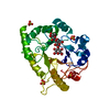
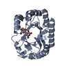
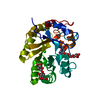
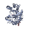
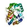
PDB-6uaq: Crystal structure of a GH128 (subgroup I) endo-beta-1,3-glucanase from Amycolatopsis mediterranei (AmGH128_I) Method: X-RAY DIFFRACTION / Resolution: 1.15 Å
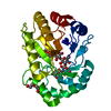
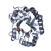
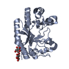
PDB-6uar: Crystal structure of a GH128 (subgroup I) endo-beta-1,3-glucanase from Amycolatopsis mediterranei (AmGH128_I) in complex with laminaritriose Method: X-RAY DIFFRACTION / Resolution: 1.4 Å
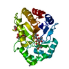
PDB-6uas: Crystal structure of a GH128 (subgroup I) endo-beta-1,3-glucanase (E199A mutant) from Amycolatopsis mediterranei (AmGH128_I) in complex with laminaripentaose Method: X-RAY DIFFRACTION / Resolution: 1.91 Å
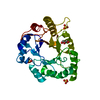
PDB-6uat: Crystal structure of a GH128 (subgroup I) endo-beta-1,3-glucanase (E102A mutant) from Amycolatopsis mediterranei (AmGH128_I) in complex with laminaripentaose Method: X-RAY DIFFRACTION / Resolution: 1.9 Å
PDB-6uau: Crystal structure of a GH128 (subgroup I) endo-beta-1,3-glucanase (E102A mutant) from Amycolatopsis mediterranei (AmGH128_I) in complex with laminaritriose and laminaribiose Method: X-RAY DIFFRACTION / Resolution: 1.9 Å
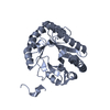
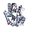
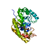
PDB-6uav: Crystal structure of a GH128 (subgroup II) endo-beta-1,3-glucanase from Pseudomonas viridiflava (PvGH128_II) Method: X-RAY DIFFRACTION / Resolution: 1.5 Å
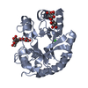
PDB-6uaw: Crystal structure of a GH128 (subgroup II) endo-beta-1,3-glucanase from Pseudomonas viridiflava (PvGH128_II) in complex with laminaritriose Method: X-RAY DIFFRACTION / Resolution: 1.5 Å
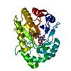
PDB-6uax: Crystal structure of a GH128 (subgroup II) endo-beta-1,3-glucanase from Sorangium cellulosum (ScGH128_II) Method: X-RAY DIFFRACTION / Resolution: 1.3 Å
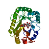
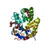
PDB-6uay: Crystal structure of a GH128 (subgroup III) curdlan-specific exo-beta-1,3-glucanase from Blastomyces gilchristii (BgGH128_III) Method: X-RAY DIFFRACTION / Resolution: 1.8 Å
PDB-6uaz: Crystal structure of a GH128 (subgroup III) curdlan-specific exo-beta-1,3-glucanase from Blastomyces gilchristii (BgGH128_III) in complex with glucose Method: X-RAY DIFFRACTION / Resolution: 1.85 Å
PDB-6ub0: Crystal structure of a GH128 (subgroup III) curdlan-specific exo-beta-1,3-glucanase from Blastomyces gilchristii (BgGH128_III) in complex with laminaribiose at -2 and -1 subsites Method: X-RAY DIFFRACTION / Resolution: 1.75 Å
PDB-6ub1: Crystal structure of a GH128 (subgroup III) curdlan-specific exo-beta-1,3-glucanase from Blastomyces gilchristii (BgGH128_III) in complex with laminaribiose at -3 and -2 subsites Method: X-RAY DIFFRACTION / Resolution: 1.6 Å
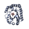
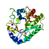
PDB-6ub2: Crystal structure of a GH128 (subgroup IV) endo-beta-1,3-glucanase from Lentinula edodes (LeGH128_IV) Method: X-RAY DIFFRACTION / Resolution: 1.8 Å
PDB-6ub3: Crystal structure of a GH128 (subgroup IV) endo-beta-1,3-glucanase from Lentinula edodes (LeGH128_IV) with laminaribiose at the surface-binding site Method: X-RAY DIFFRACTION / Resolution: 1.85 Å
PDB-6ub4: Crystal structure (C2 form) of a GH128 (subgroup IV) endo-beta-1,3-glucanase from Lentinula edodes (LeGH128_IV) in complex with laminaritriose Method: X-RAY DIFFRACTION / Resolution: 1.6 Å
PDB-6ub5: Crystal structure (P21 form) of a GH128 (subgroup IV) endo-beta-1,3-glucanase from Lentinula edodes (LeGH128_IV) in complex with laminaritriose Method: X-RAY DIFFRACTION / Resolution: 1.3 Å
PDB-6ub6: Crystal structure of a GH128 (subgroup IV) endo-beta-1,3-glucanase from Lentinula edodes (LeGH128_IV) in complex with laminaritetraose Method: X-RAY DIFFRACTION / Resolution: 1.25 Å
PDB-6ub7: Crystal structure of a GH128 (subgroup V) exo-beta-1,3-glucanase from Cryptococcus neoformans (CnGH128_V) Method: X-RAY DIFFRACTION / Resolution: 1.8 Å
PDB-6ub8: Crystal structure of a GH128 (subgroup VI) exo-beta-1,3-glucanase from Aureobasidium namibiae (AnGH128_VI) Method: X-RAY DIFFRACTION / Resolution: 1.9 Å
PDB-6uba: Crystal structure of a GH128 (subgroup VI) exo-beta-1,3-glucanase from Aureobasidium namibiae (AnGH128_VI) in complex with laminaritriose Method: X-RAY DIFFRACTION / Resolution: 2.4 Å
PDB-6ubb: Crystal structure of a GH128 (subgroup VI) exo-beta-1,3-glucanase from Aureobasidium namibiae (AnGH128_VI) with laminaribiose at the surface-binding site Method: X-RAY DIFFRACTION / Resolution: 2.35 Å
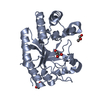
PDB-6ubc: Crystal structure of a GH128 (subgroup VII) oligosaccharide-binding protein from Cryptococcus neoformans (CnGH128_VII) Method: X-RAY DIFFRACTION / Resolution: 1.65 Å
PDB-6ubd: Crystal structure of a GH128 (subgroup VII) oligosaccharide-binding protein from Trichoderma gamsii (TgGH128_VII) Method: X-RAY DIFFRACTION / Resolution: 1.25 Å
PDB-6ufl: Crystal structure of a GH128 (subgroup I) endo-beta-1,3-glucanase (E199Q mutant) from Amycolatopsis mediterranei (AmGH128_I) in the complex with laminarihexaose Method: X-RAY DIFFRACTION / Resolution: 1.61 Å
PDB-6ufz: Crystal structure of a GH128 (subgroup I) endo-beta-1,3-glucanase (E199Q mutant) from Amycolatopsis mediterranei (AmGH128_I) Method: X-RAY DIFFRACTION / Resolution: 1.9 Å
In the structure databanks used in Yorodumi, some data are registered as the other names, "COVID-19 virus" and "2019-nCoV". Here are the details of the virus and the list of structure data.
Jan 31, 2019. EMDB accession codes are about to change! (news from PDBe EMDB page)
EMDB accession codes are about to change! (news from PDBe EMDB page)
The allocation of 4 digits for EMDB accession codes will soon come to an end. Whilst these codes will remain in use, new EMDB accession codes will include an additional digit and will expand incrementally as the available range of codes is exhausted. The current 4-digit format prefixed with “EMD-” (i.e. EMD-XXXX) will advance to a 5-digit format (i.e. EMD-XXXXX), and so on. It is currently estimated that the 4-digit codes will be depleted around Spring 2019, at which point the 5-digit format will come into force.
The EM Navigator/Yorodumi systems omit the EMD- prefix.
Related info.:Q: What is EMD? / ID/Accession-code notation in Yorodumi/EM Navigator
 Movie
Movie Controller
 Structure viewers
 About Yorodumi Papers


 Authors
 External links
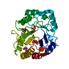


 Keywords
 HYDROLASE /
  amycolatopsis mediterranei (bacteria)
 blastomyces gilchristii (fungus)