Movie
Movie Controller
 Structure viewers
 About Yorodumi Papers


+Search query
-Structure paper
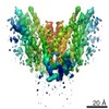
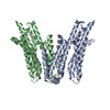
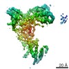
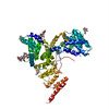
| Title | Cryo-EM structure of the protein-conducting ERAD channel Hrd1 in complex with Hrd3. |
|---|---|
| Journal, issue, pages | Nature, Vol. 548, Issue 7667, Page 352-355, Year 2017 |
| Publish date | Aug 17, 2017 |
 Authors Authors | Stefan Schoebel / Wei Mi / Alexander Stein / Sergey Ovchinnikov / Ryan Pavlovicz / Frank DiMaio / David Baker / Melissa G Chambers / Huayou Su / Dongsheng Li / Tom A Rapoport / Maofu Liao /    |
| PubMed Abstract | Misfolded endoplasmic reticulum proteins are retro-translocated through the membrane into the cytosol, where they are poly-ubiquitinated, extracted from the membrane, and degraded by the proteasome-a ...Misfolded endoplasmic reticulum proteins are retro-translocated through the membrane into the cytosol, where they are poly-ubiquitinated, extracted from the membrane, and degraded by the proteasome-a pathway termed endoplasmic reticulum-associated protein degradation (ERAD). Proteins with misfolded domains in the endoplasmic reticulum lumen or membrane are discarded through the ERAD-L and ERAD-M pathways, respectively. In Saccharomyces cerevisiae, both pathways require the ubiquitin ligase Hrd1, a multi-spanning membrane protein with a cytosolic RING finger domain. Hrd1 is the crucial membrane component for retro-translocation, but it is unclear whether it forms a protein-conducting channel. Here we present a cryo-electron microscopy structure of S. cerevisiae Hrd1 in complex with its endoplasmic reticulum luminal binding partner, Hrd3. Hrd1 forms a dimer within the membrane with one or two Hrd3 molecules associated at its luminal side. Each Hrd1 molecule has eight transmembrane segments, five of which form an aqueous cavity extending from the cytosol almost to the endoplasmic reticulum lumen, while a segment of the neighbouring Hrd1 molecule forms a lateral seal. The aqueous cavity and lateral gate are reminiscent of features of protein-conducting conduits that facilitate polypeptide movement in the opposite direction-from the cytosol into or across membranes. Our results suggest that Hrd1 forms a retro-translocation channel for the movement of misfolded polypeptides through the endoplasmic reticulum membrane. |
 External links External links | Nature / PubMed:28682307 / PubMed Central |
| Methods | EM (single particle) |
| Resolution | 3.9 - 4.7 Å |
| Structure data | EMDB-8637, PDB-5v6p: 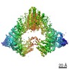 EMDB-8638: 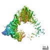 EMDB-8639: |
| Chemicals |  ChemComp-NAG: |
| Source |
|
 Keywords Keywords |  TRANSFERASE / Retrotranslocon / E3 ligase / ERAD / PROTEIN TRANSPORT / Hrd3 ERAD TRANSFERASE / Retrotranslocon / E3 ligase / ERAD / PROTEIN TRANSPORT / Hrd3 ERAD |