Movie
Movie Controller
 Structure viewers
 About Yorodumi Papers


+Search query
-Structure paper
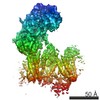
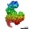
| Title | Structure of the voltage-gated calcium channel Cav1.1 complex. |
|---|---|
| Journal, issue, pages | Science, Vol. 350, Issue 6267, Page aad2395, Year 2015 |
| Publish date | Dec 18, 2015 |
 Authors Authors | Jianping Wu / Zhen Yan / Zhangqiang Li / Chuangye Yan / Shan Lu / Mengqiu Dong / Nieng Yan /  |
| PubMed Abstract | The voltage-gated calcium channel Ca(v)1.1 is engaged in the excitation-contraction coupling of skeletal muscles. The Ca(v)1.1 complex consists of the pore-forming subunit α1 and auxiliary subunits ...The voltage-gated calcium channel Ca(v)1.1 is engaged in the excitation-contraction coupling of skeletal muscles. The Ca(v)1.1 complex consists of the pore-forming subunit α1 and auxiliary subunits α2δ, β, and γ. We report the structure of the rabbit Ca(v)1.1 complex determined by single-particle cryo-electron microscopy. The four homologous repeats of the α1 subunit are arranged clockwise in the extracellular view. The γ subunit, whose structure resembles claudins, interacts with the voltage-sensing domain of repeat IV (VSD(IV)), whereas the cytosolic β subunit is located adjacent to VSD(II) of α1. The α2 subunit interacts with the extracellular loops of repeats I to III through its VWA and Cache1 domains. The structure reveals the architecture of a prototypical eukaryotic Ca(v) channel and provides a framework for understanding the function and disease mechanisms of Ca(v) and Na(v) channels. |
 External links External links | Science / PubMed:26680202 |
| Methods | EM (single particle) |
| Resolution | 4.2 - 6.1 Å |
| Structure data | |
| Chemicals |  ChemComp-CA:  ChemComp-NAG:  ChemComp-BMA: |
| Source |
|
 Keywords Keywords |  MEMBRANE PROTEIN / voltage-gated calcium channel MEMBRANE PROTEIN / voltage-gated calcium channel |