 Movie
Movie Controller
 Structure viewers
 About Yorodumi Papers


+Search query
-Structure paper
| Title | Tetrahymena RIB72A and RIB72B are microtubule inner proteins in the ciliary doublet microtubules. |
|---|---|
| Journal, issue, pages | Mol Biol Cell, Vol. 29, Issue 21, Page 2566-2577, Year 2018 |
| Publish date | Oct 15, 2018 |
 Authors Authors | Daniel Stoddard / Ying Zhao / Brian A Bayless / Long Gui / Panagiota Louka / Drashti Dave / Swati Suryawanshi / Raphaël F-X Tomasi / Pascale Dupuis-Williams / Charles N Baroud / Jacek Gaertig / Mark Winey / Daniela Nicastro /   |
| PubMed Abstract | Doublet and triplet microtubules are essential and highly stable core structures of centrioles, basal bodies, cilia, and flagella. In contrast to dynamic cytoplasmic micro-tubules, their luminal ...Doublet and triplet microtubules are essential and highly stable core structures of centrioles, basal bodies, cilia, and flagella. In contrast to dynamic cytoplasmic micro-tubules, their luminal surface is coated with regularly arranged microtubule inner proteins (MIPs). However, the protein composition and biological function(s) of MIPs remain poorly understood. Using genetic, biochemical, and imaging techniques, we identified Tetrahymena RIB72A and RIB72B proteins as ciliary MIPs. Fluorescence imaging of tagged RIB72A and RIB72B showed that both proteins colocalize to Tetrahymena cilia and basal bodies but assemble independently. Cryoelectron tomography of RIB72A and/or RIB72B knockout strains revealed major structural defects in the ciliary A-tubule involving MIP1, MIP4, and MIP6 structures. The defects of individual mutants were complementary in the double mutant. All mutants had reduced swimming speed and ciliary beat frequencies, and high-speed video imaging revealed abnormal highly curved cilia during power stroke. Our results show that RIB72A and RIB72B are crucial for the structural assembly of ciliary A-tubule MIPs and are important for proper ciliary motility. |
 External links External links | Mol Biol Cell / PubMed:30133348 / PubMed Central |
| Methods | EM (subtomogram averaging) |
| Resolution | 40.1 Å |
| Structure data |  EMDB-7805:  EMDB-7806: 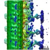 EMDB-7807:  EMDB-7811: |
| Source |
|


 Tetrahymena thermophila (eukaryote)