 Movie
Movie Controller
 Structure viewers
 About Yorodumi Papers


+Search query
-Structure paper
| Title | Structural basis of membrane bending by the N-BAR protein endophilin. |
|---|---|
| Journal, issue, pages | Cell, Vol. 149, Issue 1, Page 137-145, Year 2012 |
| Publish date | Mar 30, 2012 |
 Authors Authors | Carsten Mim / Haosheng Cui / Joseph A Gawronski-Salerno / Adam Frost / Edward Lyman / Gregory A Voth / Vinzenz M Unger /  |
| PubMed Abstract | Functioning as key players in cellular regulation of membrane curvature, BAR domain proteins bend bilayers and recruit interaction partners through poorly understood mechanisms. Using electron ...Functioning as key players in cellular regulation of membrane curvature, BAR domain proteins bend bilayers and recruit interaction partners through poorly understood mechanisms. Using electron cryomicroscopy, we present reconstructions of full-length endophilin and its N-terminal N-BAR domain in their membrane-bound state. Endophilin lattices expose large areas of membrane surface and are held together by promiscuous interactions between endophilin's amphipathic N-terminal helices. Coarse-grained molecular dynamics simulations reveal that endophilin lattices are highly dynamic and that the N-terminal helices are required for formation of a stable and regular scaffold. Furthermore, endophilin accommodates different curvatures through a quantized addition or removal of endophilin dimers, which in some cases causes dimerization of endophilin's SH3 domains, suggesting that the spatial presentation of SH3 domains, rather than affinity, governs the recruitment of downstream interaction partners. |
 External links External links | Cell / PubMed:22464326 / PubMed Central |
| Methods | EM (helical sym.) |
| Resolution | 11.0 - 26.0 Å |
| Structure data |  EMDB-2007: 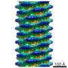 EMDB-5365: 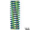 EMDB-5366:  EMDB-5367: 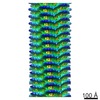 EMDB-5368: |
| Source |
|

 Rattus norvegicus (Norway rat)