 Movie
Movie Controller
 Structure viewers
 About Yorodumi Papers


+Search query
-Structure paper
| Title | Cryo-electron microscopy of the giant viruses. |
|---|---|
| Journal, issue, pages | Microscopy (Oxf), Vol. 70, Issue 6, Page 477-486, Year 2021 |
| Publish date | Nov 24, 2021 |
 Authors Authors | Raymond N Burton-Smith / Kazuyoshi Murata /  |
| PubMed Abstract | High-resolution study of the giant viruses presents one of the latest challenges in cryo-electron microscopy (EM) of viruses. Too small for light microscopy but too large for easy study at high ...High-resolution study of the giant viruses presents one of the latest challenges in cryo-electron microscopy (EM) of viruses. Too small for light microscopy but too large for easy study at high resolution by EM, they range in size from ∼0.2 to 2 μm from high-symmetry icosahedral viruses, such as Paramecium burseria Chlorella virus 1, to asymmetric forms like Tupanvirus or Pithovirus. To attain high resolution, two strategies exist to study these large viruses by cryo-EM: first, increasing the acceleration voltage of the electron microscope to improve sample penetration and overcome the limitations imposed by electro-optical physics at lower voltages, and, second, the method of 'block-based reconstruction' pioneered by Michael G. Rossmann and his collaborators, which resolves the latter limitation through an elegant leveraging of high symmetry but cannot overcome sample penetration limitations. In addition, more recent advances in both computational capacity and image processing also yield assistance in studying the giant viruses. Especially, the inclusion of Ewald sphere correction can provide large improvements in attainable resolutions for 300 kV electron microscopes. Despite this, the study of giant viruses remains a significant challenge. |
 External links External links | Microscopy (Oxf) / PubMed:34490462 |
| Methods | EM (single particle) |
| Resolution | 7.7 - 9.4 Å |
| Structure data | 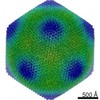 EMDB-30797: 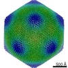 EMDB-30798: 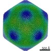 EMDB-30799: |
