Movie
Movie Controller
 Structure viewers
 About Yorodumi Papers


+Search query
-Structure paper
| Title | activates hydrolysis by recruiting and orienting on the membrane surface. |
|---|---|
| Journal, issue, pages | Proc Natl Acad Sci U S A, Vol. 120, Issue 20, Page e2301121120, Year 2023 |
| Publish date | May 16, 2023 |
 Authors Authors | Maria E Falzone / Roderick MacKinnon /  |
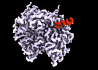
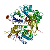
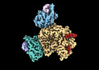
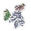
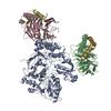
| PubMed Abstract | catalyze the hydrolysis of phosphatidylinositol 4, 5-bisphosphate [Formula: see text] into [Formula: see text] [Formula: see text] and [Formula: see text] [Formula: see text]. [Formula: see text] ... catalyze the hydrolysis of phosphatidylinositol 4, 5-bisphosphate [Formula: see text] into [Formula: see text] [Formula: see text] and [Formula: see text] [Formula: see text]. [Formula: see text] regulates the activity of many membrane proteins, while and lead to increased intracellular Ca levels and activate protein kinase C, respectively. are regulated by G protein-coupled receptors through direct interaction with [Formula: see text] and [Formula: see text] and are aqueous-soluble enzymes that must bind to the cell membrane to act on their lipid substrate. This study addresses the mechanism by which [Formula: see text] activates 3. We show that 3 functions as a slow Michaelis-Menten enzyme ( [Formula: see text] ) on membrane surfaces. We used membrane partitioning experiments to study the solution-membrane localization equilibrium of 3. Its partition coefficient is such that only a small quantity of 3 exists in the membrane in the absence of [Formula: see text] . When [Formula: see text] is present, equilibrium binding on the membrane surface increases 3 in the membrane, increasing [Formula: see text] in proportion. Atomic structures on membrane vesicle surfaces show that two [Formula: see text] anchor 3 with its catalytic site oriented toward the membrane surface. Taken together, the enzyme kinetic, membrane partitioning, and structural data show that [Formula: see text] activates by increasing its concentration on the membrane surface and orienting its catalytic core to engage [Formula: see text] . This principle of activation explains rapid stimulated catalysis with low background activity, which is essential to the biological processes mediated by [Formula: see text], and . |
 External links External links | Proc Natl Acad Sci U S A / PubMed:37172014 / PubMed Central |
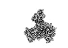
| Methods | EM (single particle) |
| Resolution | 3.3 - 3.6 Å |
| Structure data | EMDB-28266, PDB-8emv: EMDB-28267, PDB-8emw: EMDB-28268, PDB-8emx: |
| Chemicals |  ChemComp-CA: |
| Source |
|
 Keywords Keywords |  HYDROLASE / PIP2 degradation / IP3 production / DAG production / G protein signaling HYDROLASE / PIP2 degradation / IP3 production / DAG production / G protein signaling |