Movie
Movie Controller
 Structure viewers
 About Yorodumi Papers


+Search query
-Structure paper
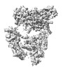
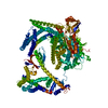
| Title | Structure of the metastatic factor P-Rex1 reveals a two-layered autoinhibitory mechanism. |
|---|---|
| Journal, issue, pages | Nat Struct Mol Biol, Vol. 29, Issue 8, Page 767-773, Year 2022 |
| Publish date | Jul 21, 2022 |
 Authors Authors | Yong-Gang Chang / Christopher J Lupton / Charles Bayly-Jones / Alastair C Keen / Laura D'Andrea / Christina M Lucato / Joel R Steele / Hari Venugopal / Ralf B Schittenhelm / James C Whisstock / Michelle L Halls / Andrew M Ellisdon /  |
| PubMed Abstract | P-Rex (PI(3,4,5)P-dependent Rac exchanger) guanine nucleotide exchange factors potently activate Rho GTPases. P-Rex guanine nucleotide exchange factors are autoinhibited, synergistically activated by ...P-Rex (PI(3,4,5)P-dependent Rac exchanger) guanine nucleotide exchange factors potently activate Rho GTPases. P-Rex guanine nucleotide exchange factors are autoinhibited, synergistically activated by Gβγ and PI(3,4,5)P binding and dysregulated in cancer. Here, we use X-ray crystallography, cryogenic electron microscopy and crosslinking mass spectrometry to determine the structural basis of human P-Rex1 autoinhibition. P-Rex1 has a bipartite structure of N- and C-terminal modules connected by a C-terminal four-helix bundle that binds the N-terminal Pleckstrin homology (PH) domain. In the N-terminal module, the Dbl homology (DH) domain catalytic surface is occluded by the compact arrangement of the DH-PH-DEP1 domains. Structural analysis reveals a remarkable conformational transition to release autoinhibition, requiring a 126° opening of the DH domain hinge helix. The off-axis position of Gβγ and PI(3,4,5)P binding sites further suggests a counter-rotation of the P-Rex1 halves by 90° facilitates PH domain uncoupling from the four-helix bundle, releasing the autoinhibited DH domain to drive Rho GTPase signaling. |
 External links External links | Nat Struct Mol Biol / PubMed:35864164 / PubMed Central |
| Methods | EM (single particle) / X-ray diffraction |
| Resolution | 3.22 - 4.4 Å |
| Structure data | EMDB-25524, PDB-7syf: 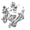 EMDB-25525: Localised reconstruction of the N-terminal half of P-Rex1 (PI(3,4,5)P3-dependent Rac Exchanger 1) 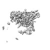 EMDB-25526: Localised reconstruction of the C-terminal half of P-Rex 1 (PI(3,4,5)P3-dependent Rac Exchanger 1) 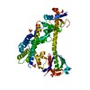 PDB-7rx9: |
| Chemicals |  ChemComp-SO4: |
| Source |
|
 Keywords Keywords |  SIGNALING PROTEIN / P-Rex1 / P-Rex2 / GEF / cell growth / Rac1 / Cdc42 / ONCOPROTEIN / guanine nucleotide exchange factor / metastasis / plasma membrane / Rho GTPase signalling SIGNALING PROTEIN / P-Rex1 / P-Rex2 / GEF / cell growth / Rac1 / Cdc42 / ONCOPROTEIN / guanine nucleotide exchange factor / metastasis / plasma membrane / Rho GTPase signalling |