 ムービー
ムービー コントローラー
 構造ビューア
 万見文献について


+検索条件
-Structure paper
| タイトル | Molecular organization of the early stages of nucleosome phase separation visualized by cryo-electron tomography. |
|---|---|
| ジャーナル・号・ページ | Mol Cell, Vol. 82, Issue 16, Page 3000-33014.e9, Year 2022 |
| 掲載日 | 2022年8月18日 |
 著者 著者 | Meng Zhang / César Díaz-Celis / Bibiana Onoa / Cristhian Cañari-Chumpitaz / Katherinne I Requejo / Jianfang Liu / Michael Vien / Eva Nogales / Gang Ren / Carlos Bustamante /  |
| PubMed 要旨 | It has been proposed that the intrinsic property of nucleosome arrays to undergo liquid-liquid phase separation (LLPS) in vitro is responsible for chromatin domain organization in vivo. However, ...It has been proposed that the intrinsic property of nucleosome arrays to undergo liquid-liquid phase separation (LLPS) in vitro is responsible for chromatin domain organization in vivo. However, understanding nucleosomal LLPS has been hindered by the challenge to characterize the structure of the resulting heterogeneous condensates. We used cryo-electron tomography and deep-learning-based 3D reconstruction/segmentation to determine the molecular organization of condensates at various stages of LLPS. We show that nucleosomal LLPS involves a two-step process: a spinodal decomposition process yielding irregular condensates, followed by their unfavorable conversion into more compact, spherical nuclei that grow into larger spherical aggregates through accretion of spinodal materials or by fusion with other spherical condensates. Histone H1 catalyzes more than 10-fold the spinodal-to-spherical conversion. We propose that this transition involves exposure of nucleosome hydrophobic surfaces causing modified inter-nucleosome interactions. These results suggest a physical mechanism by which chromatin may transition from interphase to metaphase structures. |
 リンク リンク | Mol Cell / PubMed:35907400 / PubMed Central |
| 手法 | EM (トモグラフィー) |
| 構造データ |  EMDB-24888: Molecular Organization of the Early Stages of Nucleosome Phase Separation Visualized by Cryo-Electron Tomography  EMDB-24901: Molecular Organization of the Early Stages of Nucleosome Phase Separation Visualized by Cryo-Electron Tomography 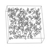 EMDB-24902: Molecular Organization of the Early Stages of Nucleosome Phase Separation Visualized by Cryo-Electron Tomography  EMDB-24903: Molecular Organization of the Early Stages of Nucleosome Phase Separation Visualized by Cryo-Electron Tomography  EMDB-24904: Molecular Organization of the Early Stages of Nucleosome Phase Separation Visualized by Cryo-Electron Tomography  EMDB-24905: Molecular Organization of the Early Stages of Nucleosome Phase Separation Visualized by Cryo-Electron Tomography  EMDB-24906: Molecular Organization of the Early Stages of Nucleosome Phase Separation Visualized by Cryo-Electron Tomography  EMDB-24907: Molecular Organization of the Early Stages of Nucleosome Phase Separation Visualized by Cryo-Electron Tomography 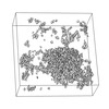 EMDB-24908: Molecular Organization of the Early Stages of Nucleosome Phase Separation Visualized by Cryo-Electron Tomography 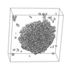 EMDB-24909: Molecular Organization of the Early Stages of Nucleosome Phase Separation Visualized by Cryo-Electron Tomography 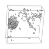 EMDB-24910: Molecular Organization of the Early Stages of Nucleosome Phase Separation Visualized by Cryo-Electron Tomography 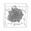 EMDB-24911: Molecular Organization of the Early Stages of Nucleosome Phase Separation Visualized by Cryo-Electron Tomography 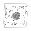 EMDB-24912: Molecular Organization of the Early Stages of Nucleosome Phase Separation Visualized by Cryo-Electron Tomography  EMDB-24913: Molecular Organization of the Early Stages of Nucleosome Phase Separation Visualized by Cryo-Electron Tomography  EMDB-24914: Molecular Organization of the Early Stages of Nucleosome Phase Separation Visualized by Cryo-Electron Tomography  EMDB-24915: Molecular Organization of the Early Stages of Nucleosome Phase Separation Visualized by Cryo-Electron Tomography  EMDB-24916: Molecular Organization of the Early Stages of Nucleosome Phase Separation Visualized by Cryo-Electron Tomography  EMDB-24917: Molecular Organization of the Early Stages of Nucleosome Phase Separation Visualized by Cryo-Electron Tomography  EMDB-24918: Molecular Organization of the Early Stages of Nucleosome Phase Separation Visualized by Cryo-Electron Tomography  EMDB-24919: Molecular Organization of the Early Stages of Nucleosome Phase Separation Visualized by Cryo-Electron Tomography  EMDB-24920: Molecular Organization of the Early Stages of Nucleosome Phase Separation Visualized by Cryo-Electron Tomography 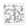 EMDB-24921: Molecular Organization of the Early Stages of Nucleosome Phase Separation Visualized by Cryo-Electron Tomography 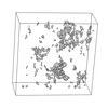 EMDB-24923: Molecular Organization of the Early Stages of Nucleosome Phase Separation Visualized by Cryo-Electron Tomography  EMDB-24924: Molecular Organization of the Early Stages of Nucleosome Phase Separation Visualized by Cryo-Electron Tomography 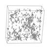 EMDB-24925: Molecular Organization of the Early Stages of Nucleosome Phase Separation Visualized by Cryo-Electron Tomography  EMDB-24926: Molecular Organization of the Early Stages of Nucleosome Phase Separation Visualized by Cryo-Electron Tomography |
| 由来 |
|
 Xenopus laevis (アフリカツメガエル)