 Movie
Movie Controller
 Structure viewers
 About Yorodumi Papers


+Search query
-Structure paper
| Title | Mechanism of membranous tunnelling nanotube formation in viral genome delivery. |
|---|---|
| Journal, issue, pages | PLoS Biol, Vol. 11, Issue 9, Page e1001667, Year 2013 |
| Publish date | Sep 24, 2013 |
 Authors Authors | Bibiana Peralta / David Gil-Carton / Daniel Castaño-Díez / Aurelie Bertin / Claire Boulogne / Hanna M Oksanen / Dennis H Bamford / Nicola G A Abrescia /  |
| PubMed Abstract | In internal membrane-containing viruses, a lipid vesicle enclosed by the icosahedral capsid protects the genome. It has been postulated that this internal membrane is the genome delivery device of ...In internal membrane-containing viruses, a lipid vesicle enclosed by the icosahedral capsid protects the genome. It has been postulated that this internal membrane is the genome delivery device of the virus. Viruses built with this architectural principle infect hosts in all three domains of cellular life. Here, using a combination of electron microscopy techniques, we investigate bacteriophage PRD1, the best understood model for such viruses, to unveil the mechanism behind the genome translocation across the cell envelope. To deliver its double-stranded DNA, the icosahedral protein-rich virus membrane transforms into a tubular structure protruding from one of the 12 vertices of the capsid. We suggest that this viral nanotube exits from the same vertex used for DNA packaging, which is biochemically distinct from the other 11. The tube crosses the capsid through an aperture corresponding to the loss of the peripentonal P3 major capsid protein trimers, penton protein P31 and membrane protein P16. The remodeling of the internal viral membrane is nucleated by changes in osmolarity and loss of capsid-membrane interactions as consequence of the de-capping of the vertices. This engages the polymerization of the tail tube, which is structured by membrane-associated proteins. We have observed that the proteo-lipidic tube in vivo can pierce the gram-negative bacterial cell envelope allowing the viral genome to be shuttled to the host cell. The internal diameter of the tube allows one double-stranded DNA chain to be translocated. We conclude that the assembly principles of the viral tunneling nanotube take advantage of proteo-lipid interactions that confer to the tail tube elastic, mechanical and functional properties employed also in other protein-membrane systems. |
 External links External links | PLoS Biol / PubMed:24086111 / PubMed Central |
| Methods | EM (subtomogram averaging) |
| Resolution | 57.0 - 66.0 Å |
| Structure data | 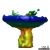 EMDB-2437:  EMDB-2438: 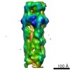 EMDB-2439: 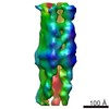 EMDB-2440: |
