Movie
Movie Controller
 Structure viewers
 About Yorodumi Papers


+Search query
-Structure paper
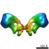
| Title | Structure of the yeast F1Fo-ATP synthase dimer and its role in shaping the mitochondrial cristae. |
|---|---|
| Journal, issue, pages | Proc Natl Acad Sci U S A, Vol. 109, Issue 34, Page 13602-13607, Year 2012 |
| Publish date | Aug 21, 2012 |
 Authors Authors | Karen M Davies / Claudio Anselmi / Ilka Wittig / José D Faraldo-Gómez / Werner Kühlbrandt /  |
| PubMed Abstract | We used electron cryotomography of mitochondrial membranes from wild-type and mutant Saccharomyces cerevisiae to investigate the structure and organization of ATP synthase dimers in situ. Subtomogram ...We used electron cryotomography of mitochondrial membranes from wild-type and mutant Saccharomyces cerevisiae to investigate the structure and organization of ATP synthase dimers in situ. Subtomogram averaging of the dimers to 3.7 nm resolution revealed a V-shaped structure of twofold symmetry, with an angle of 86° between monomers. The central and peripheral stalks are well resolved. The monomers interact within the membrane at the base of the peripheral stalks. In wild-type mitochondria ATP synthase dimers are found in rows along the highly curved cristae ridges, and appear to be crucial for membrane morphology. Strains deficient in the dimer-specific subunits e and g or the first transmembrane helix of subunit 4 lack both dimers and lamellar cristae. Instead, cristae are either absent or balloon-shaped, with ATP synthase monomers distributed randomly in the membrane. Computer simulations indicate that isolated dimers induce a plastic deformation in the lipid bilayer, which is partially relieved by their side-by-side association. We propose that the assembly of ATP synthase dimer rows is driven by the reduction in the membrane elastic energy, rather than by direct protein contacts, and that the dimer rows enable the formation of highly curved ridges in mitochondrial cristae. |
 External links External links | Proc Natl Acad Sci U S A / PubMed:22864911 / PubMed Central |
| Methods | EM (subtomogram averaging) / EM (tomography) |
| Resolution | 37.0 Å |
| Structure data | |
| Chemicals |  ChemComp-ATP:  ChemComp-MG:  ChemComp-ADP: |
| Source |
|
 Keywords Keywords |  HYDROLASE / SUBTOMOGRAM AVERAGE HYDROLASE / SUBTOMOGRAM AVERAGE |