Movie
Movie Controller
 Structure viewers
 About Yorodumi Papers


+Search query
-Structure paper
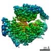
| Title | Cryo-electron microscopy structure and analysis of the P-Rex1-Gβγ signaling scaffold. |
|---|---|
| Journal, issue, pages | Sci Adv, Vol. 5, Issue 10, Page eaax8855, Year 2019 |
| Publish date | Oct 16, 2019 |
 Authors Authors | Jennifer N Cash / Sarah Urata / Sheng Li / Sandeep K Ravala / Larisa V Avramova / Michael D Shost / J Silvio Gutkind / John J G Tesmer / Michael A Cianfrocco /  |
| PubMed Abstract | PIP-dependent Rac exchanger 1 (P-Rex1) is activated downstream of G protein-coupled receptors to promote neutrophil migration and metastasis. The structure of more than half of the enzyme and its ...PIP-dependent Rac exchanger 1 (P-Rex1) is activated downstream of G protein-coupled receptors to promote neutrophil migration and metastasis. The structure of more than half of the enzyme and its regulatory G protein binding site are unknown. Our 3.2 Å cryo-EM structure of the P-Rex1-Gβγ complex reveals that the carboxyl-terminal half of P-Rex1 adopts a complex fold most similar to those of phosphoinositide phosphatases. Although catalytically inert, the domain coalesces with a DEP domain and two PDZ domains to form an extensive docking site for Gβγ. Hydrogen-deuterium exchange mass spectrometry suggests that Gβγ binding induces allosteric changes in P-Rex1, but functional assays indicate that membrane localization is also required for full activation. Thus, a multidomain assembly is key to the regulation of P-Rex1 by Gβγ and the formation of a membrane-localized scaffold optimized for recruitment of other signaling proteins such as PKA and PTEN. |
 External links External links | Sci Adv / PubMed:31663027 / PubMed Central |
| Methods | EM (single particle) |
| Resolution | 3.2 Å |
| Structure data | EMDB-20308, PDB-6pcv: |
| Source |
|
 Keywords Keywords |  SIGNALING PROTEIN / RhoGEF / G protein / Complex / Phosphatase fold SIGNALING PROTEIN / RhoGEF / G protein / Complex / Phosphatase fold |