 Movie
Movie Controller
 Structure viewers
 About Yorodumi Papers


+Search query
-Structure paper
| Title | Visualization of translation reorganization upon persistent ribosome collision stress in mammalian cells. |
|---|---|
| Journal, issue, pages | Mol Cell, Vol. 84, Issue 6, Page 1078-11089.e4, Year 2024 |
| Publish date | Mar 21, 2024 |
 Authors Authors | Juliette Fedry / Joana Silva / Mihajlo Vanevic / Stanley Fronik / Yves Mechulam / Emmanuelle Schmitt / Amédée des Georges / William James Faller / Friedrich Förster /     |
| PubMed Abstract | Aberrantly slow ribosomes incur collisions, a sentinel of stress that triggers quality control, signaling, and translation attenuation. Although each collision response has been studied in isolation, ...Aberrantly slow ribosomes incur collisions, a sentinel of stress that triggers quality control, signaling, and translation attenuation. Although each collision response has been studied in isolation, the net consequences of their collective actions in reshaping translation in cells is poorly understood. Here, we apply cryoelectron tomography to visualize the translation machinery in mammalian cells during persistent collision stress. We find that polysomes are compressed, with up to 30% of ribosomes in helical polysomes or collided disomes, some of which are bound to the stress effector GCN1. The native collision interface extends beyond the in vitro-characterized 40S and includes the L1 stalk and eEF2, possibly contributing to translocation inhibition. The accumulation of unresolved tRNA-bound 80S and 60S and aberrant 40S configurations identifies potentially limiting steps in collision responses. Our work provides a global view of the translation machinery in response to persistent collisions and a framework for quantitative analysis of translation dynamics in situ. |
 External links External links | Mol Cell / PubMed:38340715 |
| Methods | EM (subtomogram averaging) |
| Resolution | 8.8 - 40.0 Å |
| Structure data | 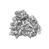 EMDB-19211: in situ subtomogram average of MEF cell ribosomes in the decoding E state  EMDB-19212: in situ subtomogram average of MEF cell ribosome in the decoding Z state 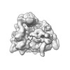 EMDB-19213: in situ subtomogram average of MEF cell ribosome in the PRE+ Z state 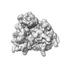 EMDB-19214: in situ subtomogram average of MEF cell ribosome in a PRE+ state 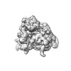 EMDB-19215: in situ subtomogram average of MEF cell ribosome in a different PRE+ state 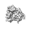 EMDB-19216: in situ subtomogram average of MEF cell ribosome in the classical PRE state 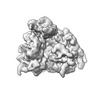 EMDB-19217: in situ subtomogram average of MEF cell ribosome in the rotated 2 state 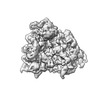 EMDB-19218: in situ subtomogram average of MEF cell ribosome in the rotated 2 + state 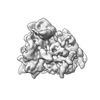 EMDB-19219: in situ subtomogram average of MEF cell ribosome in a translocation intermediate POSTi state 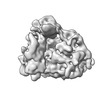 EMDB-19220: in situ subtomogram average of MEF cell ribosome in the POST state 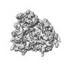 EMDB-19221: in situ subtomogram average of low dose anisomycin treated MEF cell ribosome in the OFF-P state 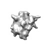 EMDB-19222: in situ subtomogram average of MEF cell pre-60S ribosome in the state B 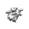 EMDB-19223: in situ subtomogram average of MEF cell idle 60S ribosome complex 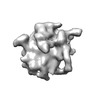 EMDB-19224: in situ subtomogram average of MEF cell ribosome associated quality control complex 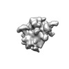 EMDB-19225: in situ subtomogram average of MEF cell non-empty 60S ribosome complex 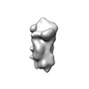 EMDB-19226: in situ subtomogram average of MEF cell 40S ribosome  EMDB-19227: in situ subtomogram average of MEF cell 48S initiation complexes 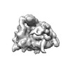 EMDB-19228: in situ subtomogram average of high dose anisomycin treated MEF cell ribosome in PRE+ Z state 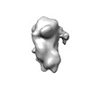 EMDB-19229: in situ subtomogram average of an aberrant 40S initiation complex in low dose anisomycin (20 min) treated MEF cell  EMDB-19230: n situ subtomogram average of aberrant initiation complex in arsenite treated MEF cells 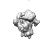 EMDB-19231: in situ subtomogram average of 43S initiation complex in low dose anisomycin treated MEF cells  EMDB-19232: in situ subtomogram average of a subclass of 43S initiation complex in low dose anisomycin treated MEF cells  EMDB-19233: in situ subtomogram average of an aberrant 40S initiation complex in low dose anisomycin treated MEF cells 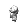 EMDB-19234: in situ subtomogram average of an aberrant 40S initiation complex in low dose anisomycin treated MEF cells 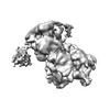 EMDB-19235: in situ subtomogram average of decoding-like stalled ribosome in low dose anisomycin treated MEF cells  EMDB-19236: in situ subtomogram average of PRE-like stalled ribosome in low dose anisomycin treated MEF cells 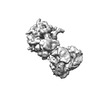 EMDB-19237: in situ subtomogram average of rotated 2 collided ribosome in low dose anisomycin treated MEF cells 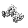 EMDB-19238: in situ subtomogram average of decoding-like collided ribosome in low dose anisomycin treated MEF cells 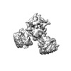 EMDB-19239: in situ subtomogram average of POSTi-like middle ribosome in helical polysomes in low dose anisomycin treated MEF cells 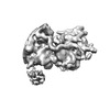 EMDB-19240: in situ subtomogram average of GCN1-bound stalled ribosome in low dose anisomycin treated MEF cells 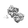 EMDB-19242: in situ subtomogram average of GCN1-bound collided ribosome in low dose anisomycin treated MEF cells |
| Source |
|

 Mus musculus (house mouse)