Yuzuru Itoh / Andreas Naschberger / Narges Mortezaei / Johannes M Herrmann / Alexey Amunts /
PubMed Abstract
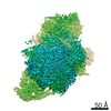
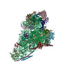
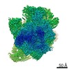
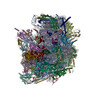
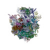
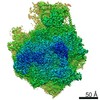
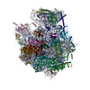
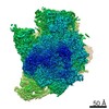
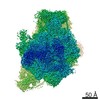
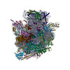
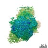
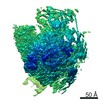
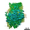
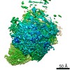
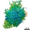
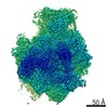
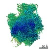
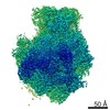
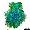
Mitoribosomes are specialized protein synthesis machineries in mitochondria. However, how mRNA binds to its dedicated channel, and tRNA moves as the mitoribosomal subunit rotate with respect to each ...Mitoribosomes are specialized protein synthesis machineries in mitochondria. However, how mRNA binds to its dedicated channel, and tRNA moves as the mitoribosomal subunit rotate with respect to each other is not understood. We report models of the translating fungal mitoribosome with mRNA, tRNA and nascent polypeptide, as well as an assembly intermediate. Nicotinamide adenine dinucleotide (NAD) is found in the central protuberance of the large subunit, and the ATPase inhibitory factor 1 (IF) in the small subunit. The models of the active mitoribosome explain how mRNA binds through a dedicated protein platform on the small subunit, tRNA is translocated with the help of the protein mL108, bridging it with L1 stalk on the large subunit, and nascent polypeptide paths through a newly shaped exit tunnel involving a series of structural rearrangements. An assembly intermediate is modeled with the maturation factor Atp25, providing insight into the biogenesis of the mitoribosomal large subunit and translation regulation.
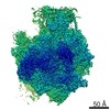
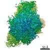
EMDB-10965, PDB-6ywe: The structure of the mitoribosome from Neurospora crassa in the P/E tRNA bound state Method: EM (single particle) / Resolution: 2.99 Å
EMDB-10966: Cryo-EM map of the core of the large subunit of the mitoribosome from Neurospora crassa with tRNA in the P/E state Method: EM (single particle) / Resolution: 2.9 Å
EMDB-10967: Cryo-EM map of the L10 region of the mitoribosome from Neurospora crassa with tRNA in the P/E state Method: EM (single particle) / Resolution: 3.14 Å
EMDB-10968: Cryo-EM map of the CP of the mitoribosome from Neurospora crassa with tRNA in the P/E state Method: EM (single particle) / Resolution: 3.12 Å
EMDB-10969: Cryo-EM map of the head of the small subunit of the mitoribosome from Neurospora crassa with tRNA in the P/E state Method: EM (single particle) / Resolution: 2.94 Å
EMDB-10970: Cryo-EM map of the body of the small subunit of the mitoribosome from Neurospora crassa with tRNA in the P/E state Method: EM (single particle) / Resolution: 2.94 Å
EMDB-10971: Cryo-EM map of the L1 stalk region of the mitoribosome from Neurospora crassa with tRNA in the P/E state Method: EM (single particle) / Resolution: 3.48 Å
EMDB-10972: Cryo-EM map of the tail region of the mitoribosome from Neurospora crassa with tRNA in the P/E state Method: EM (single particle) / Resolution: 2.99 Å
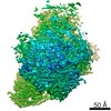
EMDB-10973, PDB-6yws: The structure of the large subunit of the mitoribosome from Neurospora crassa Method: EM (single particle) / Resolution: 2.74 Å
EMDB-10974: Cryo-EM map of the core of the large subunit of the mitoribosome from Neurospora crassa Method: EM (single particle) / Resolution: 2.7 Å
EMDB-10975: Cryo-EM map of the L10 region of the large subunit of the mitoribosome from Neurospora crassa Method: EM (single particle) / Resolution: 3.0 Å
EMDB-10976: Cryo-EM map of the CP of the large subunit of the mitoribosome from Neurospora crassa. Method: EM (single particle) / Resolution: 2.88 Å
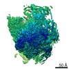
EMDB-10977, PDB-6ywv: The structure of the Atp25 bound assembly intermediate of the mitoribosome from Neurospora crassa Method: EM (single particle) / Resolution: 3.03 Å
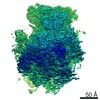
EMDB-10978, PDB-6ywx: The structure of the mitoribosome from Neurospora crassa with tRNA bound to the E-site Method: EM (single particle) / Resolution: 3.1 Å
EMDB-10979: Cryo-EM map of the core of the large subunit of the mitoribosome from Neurospora crassa with tRNA bound to the E-site Method: EM (single particle) / Resolution: 2.99 Å
EMDB-10980: Cryo-EM map of the L10 region of the mitoribosome from Neurospora crassa with tRNA bound to the E-site Method: EM (single particle) / Resolution: 3.5 Å
EMDB-10981: Cryo-EM map of the CP of the mitoribosome from Neurospora crassa Method: EM (single particle) / Resolution: 3.14 Å
EMDB-10982: Cryo-EM map of the head of the small subunit of the mitoribosome from Neurospora crassa Method: EM (single particle) / Resolution: 3.14 Å
EMDB-10983: Cryo-EM map of the tail of the small subunit of the mitoribosome from Neurospora crassa with tRNA bound to the E-site Method: EM (single particle) / Resolution: 3.21 Å
EMDB-10984: Cryo-EM map of the body of the small subunit of the mitoribosome from Neurospora crassa with tRNA bound to the E-site Method: EM (single particle) / Resolution: 3.07 Å
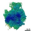
EMDB-10985, PDB-6ywy: The structure of the mitoribosome from Neurospora crassa with bound tRNA at the P-site Method: EM (single particle) / Resolution: 3.05 Å
EMDB-10986: Cryo-EM map of the core of the large subunit of the mitoribosome from Neurospora crassa with tRNA bound to the P-site Method: EM (single particle) / Resolution: 2.97 Å
EMDB-10988: Cryo-EM map of the L10 region of the mitoribosome from Neurospora crassa with tRNA bound to the P-site Method: EM (single particle) / Resolution: 3.07 Å
EMDB-10989: Cryo-EM map of the CP of the mitoribosome from Neurospora crassa with tRNA bound to the P-site Method: EM (single particle) / Resolution: 3.01 Å
EMDB-10990: Cryo-EM map of the head of the small subunit of the mitoribosome from Neurospora crassa with tRNA bound to the P-site Method: EM (single particle) / Resolution: 3.03 Å
EMDB-10991: Cryo-EM map of the body of the small subunit of the mitoribosome from Neurospora crassa with tRNA bound to the P-site Method: EM (single particle) / Resolution: 3.03 Å
EMDB-10992: Cryo-EM map for the tail of the small subunit of the mitoribosome from Neurospora crassa with tRNA bound to the P-site Method: EM (single particle) / Resolution: 3.01 Å
In the structure databanks used in Yorodumi, some data are registered as the other names, "COVID-19 virus" and "2019-nCoV". Here are the details of the virus and the list of structure data.
Jan 31, 2019. EMDB accession codes are about to change! (news from PDBe EMDB page)
EMDB accession codes are about to change! (news from PDBe EMDB page)
The allocation of 4 digits for EMDB accession codes will soon come to an end. Whilst these codes will remain in use, new EMDB accession codes will include an additional digit and will expand incrementally as the available range of codes is exhausted. The current 4-digit format prefixed with “EMD-” (i.e. EMD-XXXX) will advance to a 5-digit format (i.e. EMD-XXXXX), and so on. It is currently estimated that the 4-digit codes will be depleted around Spring 2019, at which point the 5-digit format will come into force.
The EM Navigator/Yorodumi systems omit the EMD- prefix.
Related info.:Q: What is EMD? / ID/Accession-code notation in Yorodumi/EM Navigator
 Movie
Movie Controller
 Structure viewers
 About Yorodumi Papers


 Authors

 External links
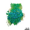


 Keywords
 TRANSLATION /