 ムービー
ムービー コントローラー
 構造ビューア
 万見文献について


+検索条件
-Structure paper
| タイトル | The 3D arrangement of the 23 S and 5 S rRNA in the Escherichia coli 50 S ribosomal subunit based on a cryo-electron microscopic reconstruction at 7.5 A resolution. |
|---|---|
| ジャーナル・号・ページ | J Mol Biol, Vol. 298, Issue 1, Page 35-59, Year 2000 |
| 掲載日 | 2000年4月21日 |
 著者 著者 | F Mueller / I Sommer / P Baranov / R Matadeen / M Stoldt / J Wöhnert / M Görlach / M van Heel / R Brimacombe /  |
| PubMed 要旨 | The Escherichia coli 23 S and 5 S rRNA molecules have been fitted helix by helix to a cryo-electron microscopic (EM) reconstruction of the 50 S ribosomal subunit, using an unfiltered version of the ...The Escherichia coli 23 S and 5 S rRNA molecules have been fitted helix by helix to a cryo-electron microscopic (EM) reconstruction of the 50 S ribosomal subunit, using an unfiltered version of the recently published 50 S reconstruction at 7.5 A resolution. At this resolution, the EM density shows a well-defined network of fine structural elements, in which the major and minor grooves of the rRNA helices can be discerned at many locations. The 3D folding of the rRNA molecules within this EM density is constrained by their well-established secondary structures, and further constraints are provided by intra and inter-rRNA crosslinking data, as well as by tertiary interactions and pseudoknots. RNA-protein cross-link and foot-print sites on the 23 S and 5 S rRNA were used to position the rRNA elements concerned in relation to the known arrangement of the ribosomal proteins as determined by immuno-electron microscopy. The published X-ray or NMR structures of seven 50 S ribosomal proteins or RNA-protein complexes were incorporated into the EM density. The 3D locations of cross-link and foot-print sites to the 23 S rRNA from tRNA bound to the ribosomal A, P or E sites were correlated with the positions of the tRNA molecules directly observed in earlier reconstructions of the 70 S ribosome at 13 A or 20 A. Similarly, the positions of cross-link sites within the peptidyl transferase ring of the 23 S rRNA from the aminoacyl residue of tRNA were correlated with the locations of the CCA ends of the A and P site tRNA. Sites on the 23 S rRNA that are cross-linked to the N termini of peptides of different lengths were all found to lie within or close to the internal tunnel connecting the peptidyl transferase region with the presumed peptide exit site on the solvent side of the 50 S subunit. The post-transcriptionally modified bases in the 23 S rRNA form a cluster close to the peptidyl transferase area. The minimum conserved core elements of the secondary structure of the 23 S rRNA form a compact block within the 3D structure and, conversely, the points corresponding to the locations of expansion segments in 28 S rRNA all lie on the outside of the structure. |
 リンク リンク | J Mol Biol / PubMed:10756104 |
| 手法 | EM (単粒子) |
| 解像度 | 7.5 Å |
| 構造データ | 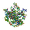 PDB-1c2w: 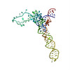 PDB-1c2x: 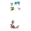 PDB-487d: |
| 由来 |
|
 キーワード キーワード |  RIBOSOME (リボソーム) / 23S RRNA (23SリボソームRNA) / LARGE RIBOSOMAL SUBUNIT / ATOMIC PROTEIN BIOSYNTHESIS / RIBONUCLEIC ACID (リボ核酸) / EM-RECONSTRUCTION / 3D ARRANGEMENT / FITTING / 5S RRNA (5SリボソームRNA) / PROTEIN BIOSYNTHESIS (タンパク質生合成) / ATOMIC STRUCTURE (原子) / RIBOSOMAL PROTEIN RIBOSOME (リボソーム) / 23S RRNA (23SリボソームRNA) / LARGE RIBOSOMAL SUBUNIT / ATOMIC PROTEIN BIOSYNTHESIS / RIBONUCLEIC ACID (リボ核酸) / EM-RECONSTRUCTION / 3D ARRANGEMENT / FITTING / 5S RRNA (5SリボソームRNA) / PROTEIN BIOSYNTHESIS (タンパク質生合成) / ATOMIC STRUCTURE (原子) / RIBOSOMAL PROTEIN |


