 ムービー
ムービー コントローラー
 構造ビューア
 万見文献について


+検索条件
-Structure paper
| タイトル | Aquaporin-0 membrane junctions reveal the structure of a closed water pore. |
|---|---|
| ジャーナル・号・ページ | Nature, Vol. 429, Issue 6988, Page 193-197, Year 2004 |
| 掲載日 | 2004年5月13日 |
 著者 著者 | Tamir Gonen / Piotr Sliz / Joerg Kistler / Yifan Cheng / Thomas Walz /  |
| PubMed 要旨 | The lens-specific water pore aquaporin-0 (AQP0) is the only aquaporin known to form membrane junctions in vivo. We show here that AQP0 from the lens core, containing some carboxy-terminally cleaved ...The lens-specific water pore aquaporin-0 (AQP0) is the only aquaporin known to form membrane junctions in vivo. We show here that AQP0 from the lens core, containing some carboxy-terminally cleaved AQP0, forms double-layered crystals that recapitulate in vivo junctions. We present the structure of the AQP0 membrane junction as determined by electron crystallography. The junction is formed by three localized interactions between AQP0 molecules in adjoining membranes, mainly mediated by proline residues conserved in AQP0s from different species but not present in most other aquaporins. Whereas all previously determined aquaporin structures show the pore in an open conformation, the water pore is closed in AQP0 junctions. The water pathway in AQP0 also contains an additional pore constriction, not seen in other known aquaporin structures, which may be responsible for pore gating. |
 リンク リンク | Nature / PubMed:15141214 |
| 手法 | EM (電子線結晶学) |
| 解像度 | 3 Å |
| 構造データ | 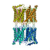 PDB-1sor: |
| 由来 |
|
 キーワード キーワード |  MEMBRANE PROTEIN (膜タンパク質) / membrane junction / water channel (アクアポリン) MEMBRANE PROTEIN (膜タンパク質) / membrane junction / water channel (アクアポリン) |

