 ムービー
ムービー コントローラー
 構造ビューア
 万見文献について


+検索条件
-Structure paper
| タイトル | Localization of L11 protein on the ribosome and elucidation of its involvement in EF-G-dependent translocation. |
|---|---|
| ジャーナル・号・ページ | J Mol Biol, Vol. 311, Issue 4, Page 777-787, Year 2001 |
| 掲載日 | 2001年8月24日 |
 著者 著者 | R K Agrawal / J Linde / J Sengupta / K H Nierhaus / J Frank /  |
| PubMed 要旨 | L11 protein is located at the base of the L7/L12 stalk of the 50 S subunit of the Escherichia coli ribosome. Because of the flexible nature of the region, recent X-ray crystallographic studies of the ...L11 protein is located at the base of the L7/L12 stalk of the 50 S subunit of the Escherichia coli ribosome. Because of the flexible nature of the region, recent X-ray crystallographic studies of the 50 S subunit failed to locate the N-terminal domain of the protein. We have determined the position of the complete L11 protein by comparing a three-dimensional cryo-EM reconstruction of the 70 S ribosome, isolated from a mutant lacking ribosomal protein L11, with the three-dimensional map of the wild-type ribosome. Fitting of the X-ray coordinates of L11-23 S RNA complex and EF-G into the cryo-EM maps combined with molecular modeling, reveals that, following EF-G-dependent GTP hydrolysis, domain V of EF-G intrudes into the cleft between the 23 S ribosomal RNA and the N-terminal domain of L11 (where the antibiotic thiostrepton binds), causing the N-terminal domain to move and thereby inducing the formation of the arc-like connection with the G' domain of EF-G. The results provide a new insight into the mechanism of EF-G-dependent translocation. |
 リンク リンク | J Mol Biol / PubMed:11518530 |
| 手法 | EM (単粒子) |
| 解像度 | 18 Å |
| 構造データ | 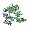 PDB-1jqm:  PDB-1jqs:  PDB-1jqt: |
| 由来 |
|
 キーワード キーワード |  RIBOSOME (リボソーム) / L11 / EF-G (EF-G) / cryo-EM (低温電子顕微鏡法) / 70s E.coli ribosome / GDP state / GTP state RIBOSOME (リボソーム) / L11 / EF-G (EF-G) / cryo-EM (低温電子顕微鏡法) / 70s E.coli ribosome / GDP state / GTP state |

