 ムービー
ムービー コントローラー
 構造ビューア
 万見文献について


+検索条件
-Structure paper
| タイトル | Defining the layers of a sensory cilium with STORM and cryoelectron nanoscopy. |
|---|---|
| ジャーナル・号・ページ | Proc Natl Acad Sci U S A, Vol. 116, Issue 47, Page 23562-23572, Year 2019 |
| 掲載日 | 2019年11月19日 |
 著者 著者 | Michael A Robichaux / Valencia L Potter / Zhixian Zhang / Feng He / Jun Liu / Michael F Schmid / Theodore G Wensel /  |
| PubMed 要旨 | Primary cilia carry out numerous signaling and sensory functions, and defects in them, "ciliopathies," cause a range of symptoms, including blindness. Understanding of their nanometer-scale ciliary ...Primary cilia carry out numerous signaling and sensory functions, and defects in them, "ciliopathies," cause a range of symptoms, including blindness. Understanding of their nanometer-scale ciliary substructures and their disruptions in ciliopathies has been hindered by limitations of conventional microscopic techniques. We have combined cryoelectron tomography, enhanced by subtomogram averaging, with superresolution stochastic optical reconstruction microscopy (STORM) to define subdomains within the light-sensing rod sensory cilium of mouse retinas and reveal previously unknown substructures formed by resident proteins. Domains are demarcated by structural features such as the axoneme and its connections to the ciliary membrane, and are correlated with molecular markers of subcompartments, including the lumen and walls of the axoneme, the membrane glycocalyx, and the intervening cytoplasm. Within this framework, we report spatial distributions of key proteins in wild-type (WT) mice and the effects on them of genetic deficiencies in 3 models of Bardet-Biedl syndrome. |
 リンク リンク | Proc Natl Acad Sci U S A / PubMed:31690665 / PubMed Central |
| 手法 | EM (トモグラフィー) |
| 構造データ | 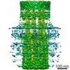 EMDB-8992: |
| 由来 |
|

 Mus musculus (ハツカネズミ)