 ムービー
ムービー コントローラー
 構造ビューア
 万見文献について


+検索条件
-Structure paper
| タイトル | Molecular structures and conformations of protocadherin-15 and its complexes on stereocilia elucidated by cryo-electron tomography. |
|---|---|
| ジャーナル・号・ページ | Elife, Vol. 10, Year 2021 |
| 掲載日 | 2021年12月29日 |
 著者 著者 | Johannes Elferich / Sarah Clark / Jingpeng Ge / April Goehring / Aya Matsui / Eric Gouaux /  |
| PubMed 要旨 | Mechanosensory transduction (MT), the conversion of mechanical stimuli into electrical signals, underpins hearing and balance and is carried out within hair cells in the inner ear. Hair cells harbor ...Mechanosensory transduction (MT), the conversion of mechanical stimuli into electrical signals, underpins hearing and balance and is carried out within hair cells in the inner ear. Hair cells harbor actin-filled stereocilia, arranged in rows of descending heights, where the tips of stereocilia are connected to their taller neighbors by a filament composed of protocadherin 15 (PCDH15) and cadherin 23 (CDH23), deemed the 'tip link.' Tension exerted on the tip link opens an ion channel at the tip of the shorter stereocilia, thus converting mechanical force into an electrical signal. While biochemical and structural studies have provided insights into the molecular composition and structure of isolated portions of the tip link, the architecture, location, and conformational states of intact tip links, on stereocilia, remains unknown. Here, we report in situ cryo-electron microscopy imaging of the tip link in mouse stereocilia. We observe individual PCDH15 molecules at the tip and shaft of stereocilia and determine their stoichiometry, conformational heterogeneity, and their complexes with other filamentous proteins, perhaps including CDH23. The PCDH15 complexes occur in clusters, frequently with more than one copy of PCDH15 at the tip of stereocilia, suggesting that tip links might consist of more than one copy of PCDH15 complexes and, by extension, might include multiple MT complexes. |
 リンク リンク | Elife / PubMed:34964715 / PubMed Central |
| 手法 | EM (トモグラフィー) |
| 構造データ |  EMDB-25046:  EMDB-25047:  EMDB-25048:  EMDB-25049:  EMDB-25050:  EMDB-25051:  EMDB-25052:  EMDB-25053:  EMDB-25054: 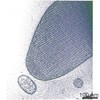 EMDB-25055: 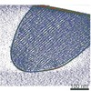 EMDB-25056: 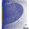 EMDB-25057: 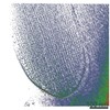 EMDB-25058:  EMDB-25059:  EMDB-25060:  EMDB-25061: |
| 由来 |
|

 Oryctolagus cuniculus (ウサギ)