 ムービー
ムービー コントローラー
 構造ビューア
 万見文献について


+検索条件
-Structure paper
| タイトル | The 3-dimensional structure of a hepatitis C virus p7 ion channel by electron microscopy. |
|---|---|
| ジャーナル・号・ページ | Proc Natl Acad Sci U S A, Vol. 106, Issue 31, Page 12712-12716, Year 2009 |
| 掲載日 | 2009年8月4日 |
 著者 著者 | Philipp Luik / Chee Chew / Jussi Aittoniemi / Jason Chang / Paul Wentworth / Raymond A Dwek / Philip C Biggin / Catherine Vénien-Bryan / Nicole Zitzmann /  |
| PubMed 要旨 | Infection with the hepatitis C virus (HCV) has a huge impact on global health putting more than 170 million people at risk of developing severe liver disease. The HCV encoded p7 ion channel is ...Infection with the hepatitis C virus (HCV) has a huge impact on global health putting more than 170 million people at risk of developing severe liver disease. The HCV encoded p7 ion channel is essential for the production of infectious viruses. Despite a growing body of functional data, little is known about the 3-dimensional (3D) structure of the channel. Here, we present the 3D structure of a full-length viroporin, the detergent-solubilized hexameric 42 kDa form of the HCV p7 ion channel, as determined by single-particle electron microscopy using the random conical tilting approach. The reconstruction of such a small protein complex was made possible by a combination of high-contrast staining, the symmetry, and the distinct structural features of the channel. The orientation of the p7 monomers within the density was established using immunolabeling with N and C termini specific F(ab) fragments. The density map at a resolution of approximately 16 A reveals a flower-shaped protein architecture with protruding petals oriented toward the ER lumen. This broadest part of the channel presents a comparatively large surface area providing potential interaction sites for cellular and virally encoded ER resident proteins. |
 リンク リンク | Proc Natl Acad Sci U S A / PubMed:19590017 / PubMed Central |
| 手法 | EM (単粒子) |
| 解像度 | 16.0 Å |
| 構造データ | 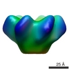 EMDB-1661: |
| 由来 |
|
 Hepatitis C virus (ウイルス)