 ムービー
ムービー コントローラー
 構造ビューア
 万見文献について


+検索条件
-Structure paper
| タイトル | The Hantavirus Surface Glycoprotein Lattice and Its Fusion Control Mechanism. |
|---|---|
| ジャーナル・号・ページ | Cell, Vol. 183, Issue 2, Page 442-456.e16, Year 2020 |
| 掲載日 | 2020年10月15日 |
 著者 著者 | Alexandra Serris / Robert Stass / Eduardo A Bignon / Nicolás A Muena / Jean-Claude Manuguerra / Rohit K Jangra / Sai Li / Kartik Chandran / Nicole D Tischler / Juha T Huiskonen / Felix A Rey / Pablo Guardado-Calvo /       |
| PubMed 要旨 | Hantaviruses are rodent-borne viruses causing serious zoonotic outbreaks worldwide for which no treatment is available. Hantavirus particles are pleomorphic and display a characteristic square ...Hantaviruses are rodent-borne viruses causing serious zoonotic outbreaks worldwide for which no treatment is available. Hantavirus particles are pleomorphic and display a characteristic square surface lattice. The envelope glycoproteins Gn and Gc form heterodimers that further assemble into tetrameric spikes, the lattice building blocks. The glycoproteins, which are the sole targets of neutralizing antibodies, drive virus entry via receptor-mediated endocytosis and endosomal membrane fusion. Here we describe the high-resolution X-ray structures of the heterodimer of Gc and the Gn head and of the homotetrameric Gn base. Docking them into an 11.4-Å-resolution cryoelectron tomography map of the hantavirus surface accounted for the complete extramembrane portion of the viral glycoprotein shell and allowed a detailed description of the surface organization of these pleomorphic virions. Our results, which further revealed a built-in mechanism controlling Gc membrane insertion for fusion, pave the way for immunogen design to protect against pathogenic hantaviruses. |
 リンク リンク | Cell / PubMed:32937107 / PubMed Central |
| 手法 | EM (サブトモグラム平均) / X線回折 |
| 解像度 | 1.902 - 15.6 Å |
| 構造データ | EMDB-11236: Reconstruction of Tula virus surface glycoprotein lattice 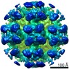 EMDB-4867:  PDB-6y5f:  PDB-6y5w:  PDB-6y62:  PDB-6y68:  PDB-6y6p:  PDB-6y6q:  PDB-6yrb:  PDB-6yrq: |
| 化合物 |  ChemComp-NAG:  ChemComp-PO4:  ChemComp-HOH:  ChemComp-CD:  ChemComp-SO4:  ChemComp-IOD:  ChemComp-MPD: |
| 由来 |
|
 キーワード キーワード |  VIRAL PROTEIN (ウイルスタンパク質) / class-II fusion protein hantavirus bunyavirus prefusion complex / class-II fusion protein hantavirus bunyavirus / prefusion complex / postfusion conformation / Hantavirus (ハンタウイルス) / Tula / Andes / TULV / ANDV / glycoprotein (糖タンパク質) / lattice / spike / fusion protein (融合タンパク質) VIRAL PROTEIN (ウイルスタンパク質) / class-II fusion protein hantavirus bunyavirus prefusion complex / class-II fusion protein hantavirus bunyavirus / prefusion complex / postfusion conformation / Hantavirus (ハンタウイルス) / Tula / Andes / TULV / ANDV / glycoprotein (糖タンパク質) / lattice / spike / fusion protein (融合タンパク質) |


 tula orthohantavirus (ウイルス)
