 ムービー
ムービー コントローラー
 構造ビューア
 万見文献について


+検索条件
-Structure paper
| タイトル | Origin and arrangement of actin filaments for gliding motility in apicomplexan parasites revealed by cryo-electron tomography. |
|---|---|
| ジャーナル・号・ページ | Nat Commun, Vol. 14, Issue 1, Page 4800, Year 2023 |
| 掲載日 | 2023年8月9日 |
 著者 著者 | Matthew Martinez / Shrawan Kumar Mageswaran / Amandine Guérin / William David Chen / Cameron Parker Thompson / Sabine Chavin / Dominique Soldati-Favre / Boris Striepen / Yi-Wei Chang /   |
| PubMed 要旨 | The phylum Apicomplexa comprises important eukaryotic parasites that invade host tissues and cells using a unique mechanism of gliding motility. Gliding is powered by actomyosin motors that ...The phylum Apicomplexa comprises important eukaryotic parasites that invade host tissues and cells using a unique mechanism of gliding motility. Gliding is powered by actomyosin motors that translocate host-attached surface adhesins along the parasite cell body. Actin filaments (F-actin) generated by Formin1 play a central role in this critical parasitic activity. However, their subcellular origin, path and ultrastructural arrangement are poorly understood. Here we used cryo-electron tomography to image motile Cryptosporidium parvum sporozoites and reveal the cellular architecture of F-actin at nanometer-scale resolution. We demonstrate that F-actin nucleates at the apically positioned preconoidal rings and is channeled into the pellicular space between the parasite plasma membrane and the inner membrane complex in a conoid extrusion-dependent manner. Within the pellicular space, filaments on the inner membrane complex surface appear to guide the apico-basal flux of F-actin. F-actin concordantly accumulates at the basal end of the parasite. Finally, analyzing a Formin1-depleted Toxoplasma gondii mutant pinpoints the upper preconoidal ring as the conserved nucleation hub for F-actin in Cryptosporidium and Toxoplasma. Together, we provide an ultrastructural model for the life cycle of F-actin for apicomplexan gliding motility. |
 リンク リンク | Nat Commun / PubMed:37558667 / PubMed Central |
| 手法 | EM (トモグラフィー) / EM (サブトモグラム平均) |
| 解像度 | 30.0 - 65.0 Å |
| 構造データ | 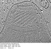 EMDB-29753: Tomogram of an apical end of a Toxoplasma Gondii tachyzoite displaying an extruded conoid 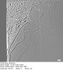 EMDB-29754: Cryptosporidium parvum sporozoite apical end 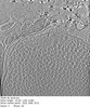 EMDB-29755: Cryptosporidium parvum sporozoite basal end  EMDB-29784: Subtomogram average of the preconoidal rings from Cryptosporidium parvum sporozoites 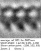 EMDB-29791: Refined subtomogram average of the top preconoidal ring from Cryptosporidium parvum sporozoites  EMDB-29801: Refined subtomogram average of the bottom preconoidal ring from Cryptosporidium parvum sporozoites  EMDB-29808: Subtomogram average of the IMC surface filaments, top view, from Cryptosporidium parvum sporozoites  EMDB-29809: IMC surface filament, sawtooth conformation, from Cryptosporidium parvum sporozoites  EMDB-29810: IMC surface filament, side view, from Cryptosporidium parvum sporozoites  EMDB-29826: Upper preconoidal ring from Toxoplasma gondii tachyzoites  EMDB-29827: Lower preconoidal ring of Toxoplasma gondii tachyzoites  EMDB-29832: Preconoidal rings of Toxoplasma gondii tachyzoites  EMDB-29835: Basal IMC pore of Cryptosporidium parvum sporozoites  EMDB-29838: Preconoidal rings of Toxoplasma gondii tachyzoites with Formin1 conditionally depleted  EMDB-29839: Upper preconoidal ring of Toxoplasma gondii tachyzoites with Formin1 conditionally depleted  EMDB-29840: Lower preconoidal ring from Toxoplasma gondii tachyzoites with Formin1 conditionally depleted |
| 由来 |
|

 Toxoplasma gondii (トキソプラズマ)