 Movie
Movie Controller
 Structure viewers
 About Yorodumi Papers


+Search query
-Structure paper
| Title | Key Intermediates in Ribosome Recycling Visualized by Time-Resolved Cryoelectron Microscopy. |
|---|---|
| Journal, issue, pages | Structure, Vol. 24, Issue 12, Page 2092-2101, Year 2016 |
| Publish date | Dec 6, 2016 |
 Authors Authors | Ziao Fu / Sandip Kaledhonkar / Anneli Borg / Ming Sun / Bo Chen / Robert A Grassucci / Måns Ehrenberg / Joachim Frank /   |
| PubMed Abstract | Upon encountering a stop codon on mRNA, polypeptide synthesis on the ribosome is terminated by release factors, and the ribosome complex, still bound with mRNA and P-site-bound tRNA (post-termination ...Upon encountering a stop codon on mRNA, polypeptide synthesis on the ribosome is terminated by release factors, and the ribosome complex, still bound with mRNA and P-site-bound tRNA (post-termination complex, PostTC), is split into ribosomal subunits, ready for a new round of translational initiation. Separation of post-termination ribosomes into subunits, or "ribosome recycling," is promoted by the joint action of ribosome-recycling factor (RRF) and elongation factor G (EF-G) in a guanosine triphosphate (GTP) hydrolysis-dependent manner. Here we used a mixing-spraying-based method of time-resolved cryo-electron microscopy (cryo-EM) to visualize the short-lived intermediates of the recycling process. The two complexes that contain (1) both RRF and EF-G bound to the PostTC or (2) deacylated tRNA bound to the 30S subunit are of particular interest. Our observations of the native form of these complexes demonstrate the strong potential of time-resolved cryo-EM for visualizing previously unobservable transient structures. |
 External links External links | Structure / PubMed:27818103 / PubMed Central |
| Methods | EM (single particle) |
| Resolution | 7.4 - 16.0 Å |
| Structure data | 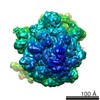 EMDB-8411: 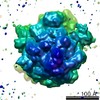 EMDB-8412: 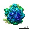 EMDB-8413: 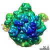 EMDB-8415: 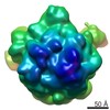 EMDB-8416: 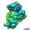 EMDB-8417: 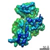 EMDB-8418: |
| Source |
|

 Escherichia coli (E. coli)