Movie
Movie Controller
 Structure viewers
 About Yorodumi Papers


+Search query
-Structure paper
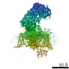
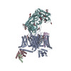
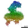
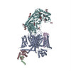
| Title | Structure of the voltage-gated calcium channel Ca(v)1.1 at 3.6 Å resolution. |
|---|---|
| Journal, issue, pages | Nature, Vol. 537, Issue 7619, Page 191-196, Year 2016 |
| Publish date | Sep 8, 2016 |
 Authors Authors | Jianping Wu / Zhen Yan / Zhangqiang Li / Xingyang Qian / Shan Lu / Mengqiu Dong / Qiang Zhou / Nieng Yan /  |
| PubMed Abstract | The voltage-gated calcium (Ca) channels convert membrane electrical signals to intracellular Ca-mediated events. Among the ten subtypes of Ca channel in mammals, Ca1.1 is specified for the excitation- ...The voltage-gated calcium (Ca) channels convert membrane electrical signals to intracellular Ca-mediated events. Among the ten subtypes of Ca channel in mammals, Ca1.1 is specified for the excitation-contraction coupling of skeletal muscles. Here we present the cryo-electron microscopy structure of the rabbit Ca1.1 complex at a nominal resolution of 3.6 Å. The inner gate of the ion-conducting α1-subunit is closed and all four voltage-sensing domains adopt an 'up' conformation, suggesting a potentially inactivated state. The extended extracellular loops of the pore domain, which are stabilized by multiple disulfide bonds, form a windowed dome above the selectivity filter. One side of the dome provides the docking site for the α2δ-1-subunit, while the other side may attract cations through its negative surface potential. The intracellular I-II and III-IV linker helices interact with the β-subunit and the carboxy-terminal domain of α1, respectively. Classification of the particles yielded two additional reconstructions that reveal pronounced displacement of β and adjacent elements in α1. The atomic model of the Ca1.1 complex establishes a foundation for mechanistic understanding of excitation-contraction coupling and provides a three-dimensional template for molecular interpretations of the functions and disease mechanisms of Ca and Na channels. |
 External links External links | Nature / PubMed:27580036 |
| Methods | EM (single particle) |
| Resolution | 3.6 - 3.9 Å |
| Structure data | EMDB-9513: Cryo-EM map of the rabbit voltage-gated calcium channel Cav1.1 at 3.6 angstrom resolution 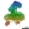 EMDB-9514: |
| Chemicals |  ChemComp-NAG:  ChemComp-PC1:  ChemComp-CA: |
| Source |
|
 Keywords Keywords |  MEMBRANE PROTEIN / complex / channel MEMBRANE PROTEIN / complex / channel |