Movie
Movie Controller
 Structure viewers
 About Yorodumi Papers


+Search query
-Structure paper
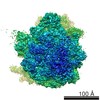
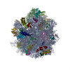
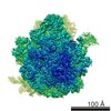
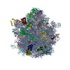
| Title | Structures of ribosome-bound initiation factor 2 reveal the mechanism of subunit association. |
|---|---|
| Journal, issue, pages | Sci Adv, Vol. 2, Issue 3, Page e1501502, Year 2016 |
| Publish date | Mar 4, 2016 |
 Authors Authors | Thiemo Sprink / David J F Ramrath / Hiroshi Yamamoto / Kaori Yamamoto / Justus Loerke / Jochen Ismer / Peter W Hildebrand / Patrick Scheerer / Jörg Bürger / Thorsten Mielke / Christian M T Spahn /  |
| PubMed Abstract | Throughout the four phases of protein biosynthesis-initiation, elongation, termination, and recycling-the ribosome is controlled and regulated by at least one specified translational guanosine ...Throughout the four phases of protein biosynthesis-initiation, elongation, termination, and recycling-the ribosome is controlled and regulated by at least one specified translational guanosine triphosphatase (trGTPase). Although the structural basis for trGTPase interaction with the ribosome has been solved for the last three steps of translation, the high-resolution structure for the key initiation trGTPase, initiation factor 2 (IF2), complexed with the ribosome, remains elusive. We determine the structure of IF2 complexed with a nonhydrolyzable guanosine triphosphate analog and initiator fMet-tRNAi (Met) in the context of the Escherichia coli ribosome to 3.7-Å resolution using cryo-electron microscopy. The structural analysis reveals previously unseen intrinsic conformational modes of the 70S initiation complex, establishing the mutual interplay of IF2 and initator transfer RNA (tRNA) with the ribsosome and providing the structural foundation for a mechanistic understanding of the final steps of translation initiation. |
 External links External links | Sci Adv / PubMed:26973877 / PubMed Central |
| Methods | EM (single particle) |
| Resolution | 3.7 - 4.6 Å |
| Structure data | |
| Chemicals |  ChemComp-MG:  ChemComp-ZN:  ChemComp-GNP:  ChemComp-FME:  ChemComp-HOH: |
| Source |
|
 Keywords Keywords |  RIBOSOME / translation initiation / translation initiation factor 2 / IF2 / translational GTPase / 70S / fMet-tRNA / GTPase / bacterial ribosome / initiation factor 2 RIBOSOME / translation initiation / translation initiation factor 2 / IF2 / translational GTPase / 70S / fMet-tRNA / GTPase / bacterial ribosome / initiation factor 2 |