 Movie
Movie Controller
 Structure viewers
 About Yorodumi Papers


+Search query
-Structure paper
| Title | Electron crystallography of ultrathin 3D protein crystals: atomic model with charges. |
|---|---|
| Journal, issue, pages | Proc Natl Acad Sci U S A, Vol. 112, Issue 11, Page 3368-3373, Year 2015 |
| Publish date | Mar 17, 2015 |
 Authors Authors | Koji Yonekura / Kazuyuki Kato / Mitsuo Ogasawara / Masahiro Tomita / Chikashi Toyoshima /  |
| PubMed Abstract | Membrane proteins and macromolecular complexes often yield crystals too small or too thin for even the modern synchrotron X-ray beam. Electron crystallography could provide a powerful means for ...Membrane proteins and macromolecular complexes often yield crystals too small or too thin for even the modern synchrotron X-ray beam. Electron crystallography could provide a powerful means for structure determination with such undersized crystals, as protein atoms diffract electrons four to five orders of magnitude more strongly than they do X-rays. Furthermore, as electron crystallography yields Coulomb potential maps rather than electron density maps, it could provide a unique method to visualize the charged states of amino acid residues and metals. Here we describe an attempt to develop a methodology for electron crystallography of ultrathin (only a few layers thick) 3D protein crystals and present the Coulomb potential maps at 3.4-Å and 3.2-Å resolution, respectively, obtained from Ca(2+)-ATPase and catalase crystals. These maps demonstrate that it is indeed possible to build atomic models from such crystals and even to determine the charged states of amino acid residues in the Ca(2+)-binding sites of Ca(2+)-ATPase and that of the iron atom in the heme in catalase. |
 External links External links | Proc Natl Acad Sci U S A / PubMed:25730881 / PubMed Central |
| Methods | EM (electron crystallography) |
| Resolution | 3.2 - 3.4 Å |
| Structure data | 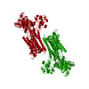 PDB-3j7t: 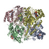 PDB-5gkn: |
| Chemicals |  ChemComp-CA:  ChemComp-NA:  ChemComp-HOH:  ChemComp-HEM:  ChemComp-NDP: |
| Source |
|
 Keywords Keywords |  HYDROLASE / ION PUMP / MEMBRANE PROTEIN / P-TYPE ATPASE / ACTIVE TRANSPORT / OXIDOREDUCTASE / HEME / NADPH HYDROLASE / ION PUMP / MEMBRANE PROTEIN / P-TYPE ATPASE / ACTIVE TRANSPORT / OXIDOREDUCTASE / HEME / NADPH |

