 Movie
Movie Controller
 Structure viewers
 About Yorodumi Papers


+Search query
-Structure paper
| Title | Yeast rvb1 and rvb2 proteins oligomerize as a conformationally variable dodecamer with low frequency. |
|---|---|
| Journal, issue, pages | J Mol Biol, Vol. 427, Issue 10, Page 1875-1886, Year 2015 |
| Publish date | May 22, 2015 |
 Authors Authors | Ajitha Jeganathan / Vivian Leong / Liang Zhao / Jennifer Huen / Nardin Nano / Walid A Houry / Joaquin Ortega /  |
| PubMed Abstract | Rvb1 and Rvb2 are conserved AAA+ (ATPases associated with diverse cellular activities) proteins found at the core of large multicomponent complexes that play key roles in chromatin remodeling, ...Rvb1 and Rvb2 are conserved AAA+ (ATPases associated with diverse cellular activities) proteins found at the core of large multicomponent complexes that play key roles in chromatin remodeling, integrity of the telomeres, ribonucleoprotein complex biogenesis and other essential cellular processes. These proteins contain an AAA+ domain for ATP binding and hydrolysis and an insertion domain proposed to bind DNA/RNA. Yeast Rvb1 and Rvb2 proteins oligomerize primarily as heterohexameric rings. The six AAA+ core domains form the body of the ring and the insertion domains protrude from one face of the ring. Conversely, human Rvbs form a mixture of hexamers and dodecamers made of two stacked hexamers interacting through the insertion domains. Human dodecamers adopt either a contracted or a stretched conformation. Here, we found that yeast Rvb1/Rvb2 complexes when assembled in vivo mainly form hexamers but they also assemble as dodecamers with a frequency lower than 10%. Yeast dodecamers adopt not only the stretched and contracted structures that have been described for human Rvb1/Rvb2 dodecamers but also intermediate conformations in between these two extreme states. The orientation of the insertion domains of Rvb1 and Rvb2 proteins in these conformers changes as the dodecamer transitions from the stretched structure to a more contracted structure. Finally, we observed that for the yeast proteins, oligomerization as a dodecamer inhibits the ATPase activity of the Rvb1/Rvb2 complex. These results indicate that although human and yeast Rvb1 and Rvb2 proteins share high degree of homology, there are significant differences in their oligomeric behavior and dynamics. |
 External links External links | J Mol Biol / PubMed:25636407 |
| Methods | EM (single particle) |
| Resolution | 25.0 Å |
| Structure data | 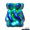 EMDB-6215:  EMDB-6216:  EMDB-6217: 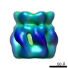 EMDB-6218: |
| Source |
|

 Saccharomyces cerevisiae (brewer's yeast)