 Movie
Movie Controller
 Structure viewers
 About Yorodumi Papers


+Search query
-Structure paper
| Title | A structural model of the genome packaging process in a membrane-containing double stranded DNA virus. |
|---|---|
| Journal, issue, pages | PLoS Biol, Vol. 12, Issue 12, Page e1002024, Year 2014 |
| Publish date | Dec 16, 2014 |
 Authors Authors | Chuan Hong / Hanna M Oksanen / Xiangan Liu / Joanita Jakana / Dennis H Bamford / Wah Chiu /   |
| PubMed Abstract | Two crucial steps in the virus life cycle are genome encapsidation to form an infective virion and genome exit to infect the next host cell. In most icosahedral double-stranded (ds) DNA viruses, the ...Two crucial steps in the virus life cycle are genome encapsidation to form an infective virion and genome exit to infect the next host cell. In most icosahedral double-stranded (ds) DNA viruses, the viral genome enters and exits the capsid through a unique vertex. Internal membrane-containing viruses possess additional complexity as the genome must be translocated through the viral membrane bilayer. Here, we report the structure of the genome packaging complex with a membrane conduit essential for viral genome encapsidation in the tailless icosahedral membrane-containing bacteriophage PRD1. We utilize single particle electron cryo-microscopy (cryo-EM) and symmetry-free image reconstruction to determine structures of PRD1 virion, procapsid, and packaging deficient mutant particles. At the unique vertex of PRD1, the packaging complex replaces the regular 5-fold structure and crosses the lipid bilayer. These structures reveal that the packaging ATPase P9 and the packaging efficiency factor P6 form a dodecameric portal complex external to the membrane moiety, surrounded by ten major capsid protein P3 trimers. The viral transmembrane density at the special vertex is assigned to be a hexamer of heterodimer of proteins P20 and P22. The hexamer functions as a membrane conduit for the DNA and as a nucleating site for the unique vertex assembly. Our structures show a conformational alteration in the lipid membrane after the P9 and P6 are recruited to the virion. The P8-genome complex is then packaged into the procapsid through the unique vertex while the genome terminal protein P8 functions as a valve that closes the channel once the genome is inside. Comparing mature virion, procapsid, and mutant particle structures led us to propose an assembly pathway for the genome packaging apparatus in the PRD1 virion. |
 External links External links | PLoS Biol / PubMed:25514469 / PubMed Central |
| Methods | EM (single particle) |
| Resolution | 12.0 - 22.0 Å |
| Structure data | 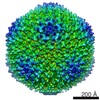 EMDB-5984: 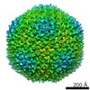 EMDB-5985: 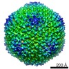 EMDB-5986: 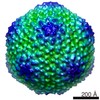 EMDB-5987: 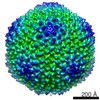 EMDB-5988: |
